Product Description
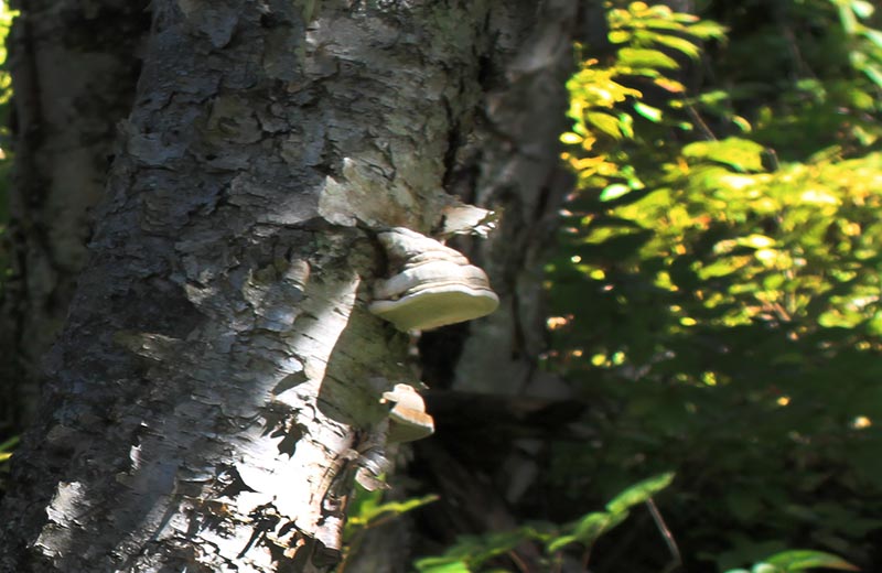
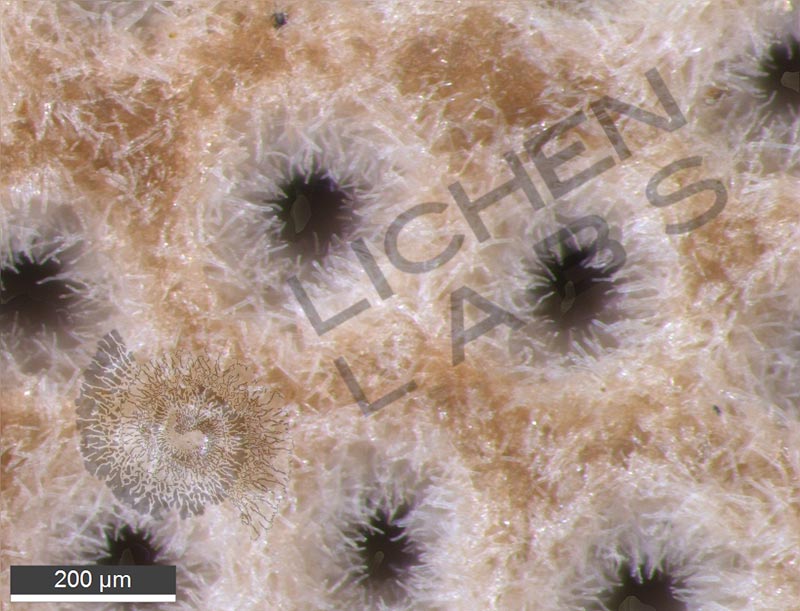
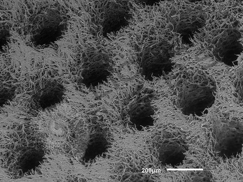
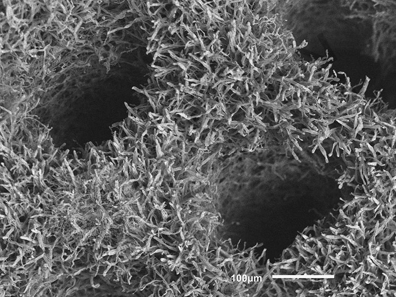
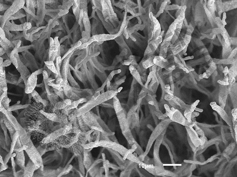
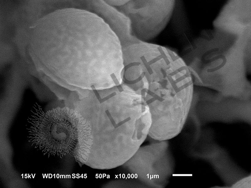
Bracket fungus can grow in wounds on the bark on living trees or on the bark of older or dead trees. The fungus decays the wood and produces a perennial “mushroom” fruiting body. Yearly growth occurs on the bottom of the fungus, and it can live up to 30 years. It has a woody fibrous structure with long round pores that will bear spores appearing as a white powder.
There are 2 types in the north woods. The first is Birch polypore (Fomitopsis betulina), shown on a birch tree in the macro product picture. It causes brown rot on birch trees and logs. It is known to have many medicinal effects, including anti-bacterial, anti-fungal, and anti-virus properties. It was one of the mushrooms found with the “Tyrolean Iceman,” a 5000-year-old mummified body discovered in the Italian Alps in 1991. He appeared to be carrying it in his “first aid kit.” The body was carbon-dated to between 3350 and 3100 BC. https://icemanotzi.weebly.com/artifacts.html and https://link.springer.com/content/pdf/10.1007/s11274-017-2247-0.pdf
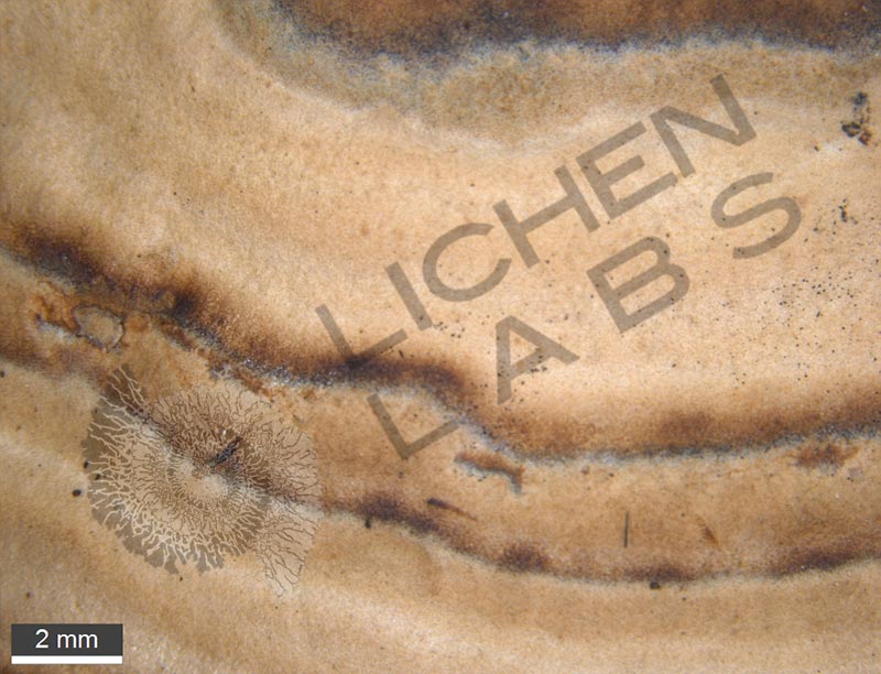
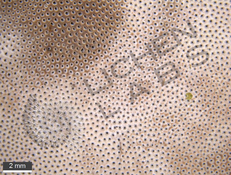
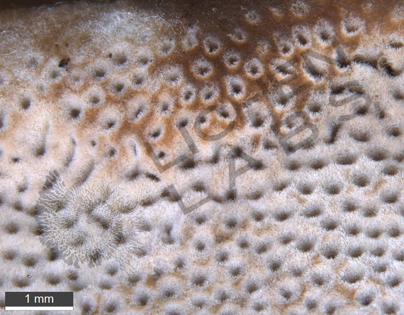
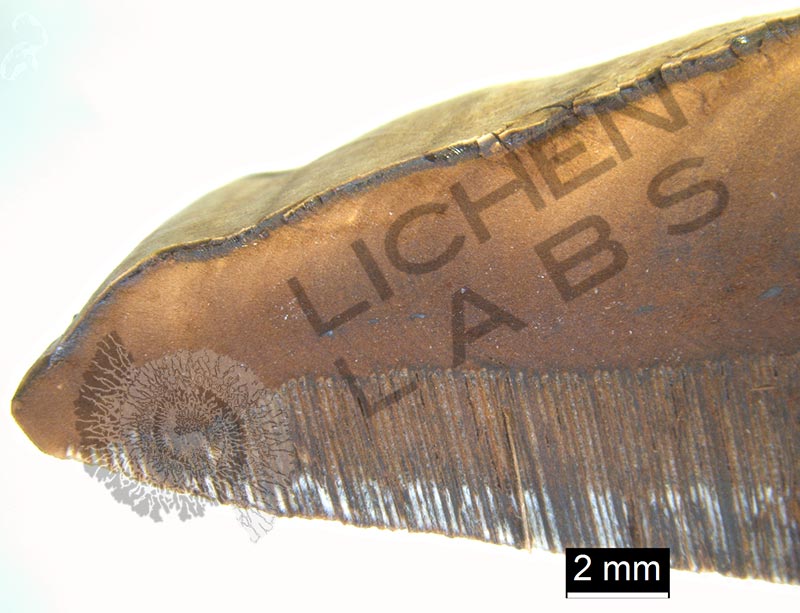
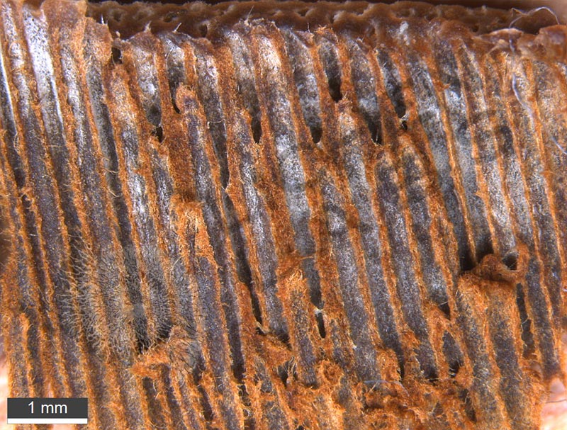
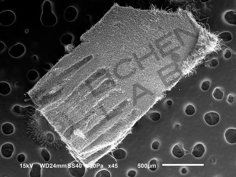
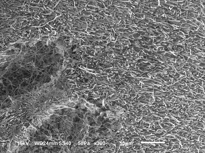
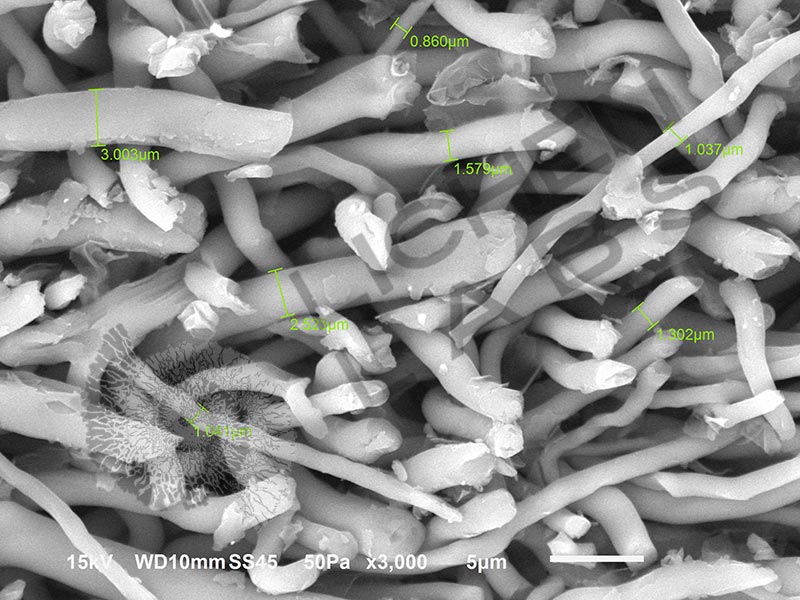
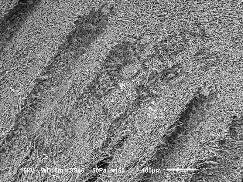
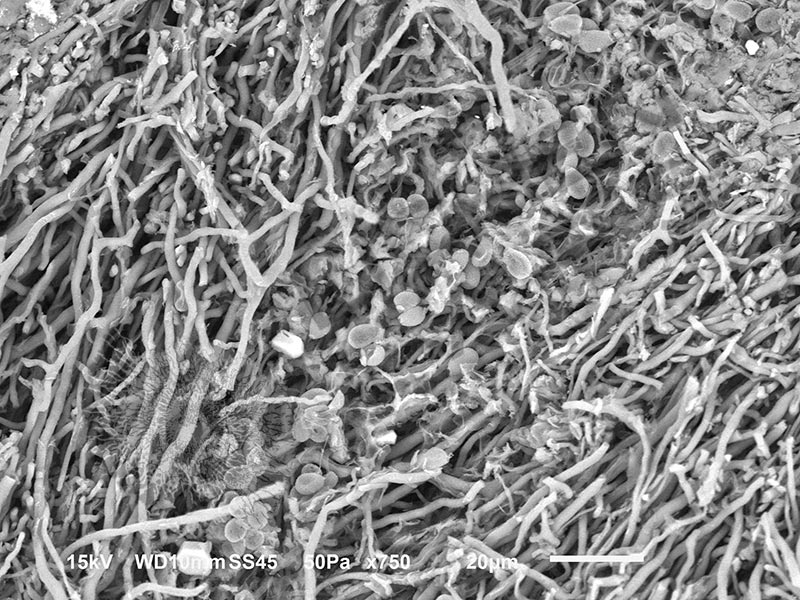
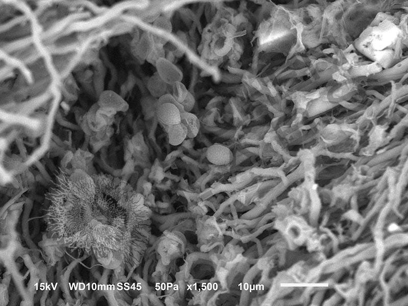
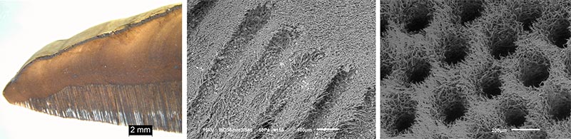
The other species, Tinder polypore (Formes fomentarius) is banded on the top side. It causes white rot and also grows on birch as well as willow, poplar, aspen and other deciduous trees. It has a woody fibrous structure with long round pores shown in the cross-section above left and center and from the bottom of the fungus at right. It what will bear spores that appear as a white powder. In addition to medicinal uses, it was also consumed as tinder for starting fires.
PURCHASE 5 OR MORE IMAGES AND GET 20% OFF YOUR ENTIRE ORDER!
Please contact us for custom images.
The image store is a collection of organisms that have been examined under a stereo light microscope (LM) and or scanning electron microscope (SEM). Each group of organisms has a short description and a longer more detailed description or story about the organism. Clicking on the product group shows the individual images. Each series takes the observer from macro to micro or nano on a particular organism, starting with a macro photographic image(s) for perspective, micro images taken by the light microscope, and most have micro to nano scanning electron microscope images. The SEM images will appear in black and white as a beam of electrons is used to illuminate the specimen rather than light. A few SEM images are colorized (lotus leaf). More information about the labeling and techniques used is below.
For the curious:
The light microscope images are labeled LM and a Z is included if it is a vertical composite of images effectively extending the depth of field or EDF of the microscope.
SEM images are labeled by the type of detector use:
SE (secondary electron)
LSE (Low vacuum secondary electron)
BES (backscattered electron shadow mode)
BEC (backscattered electron compositional mode)
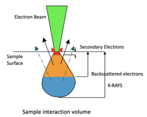
The SEM instrument works by producing a beam of electrons under a vacuum that interacts with the sample surface and subsurface producing different signals, as shown in the diagram at right. Secondary electrons, backscattered electrons and x-rays are detected using different instrument modes. In addition to morphological information to produce an image the SEM can determine elemental composition by energy dispersive x-ray spectroscopy (EDS).