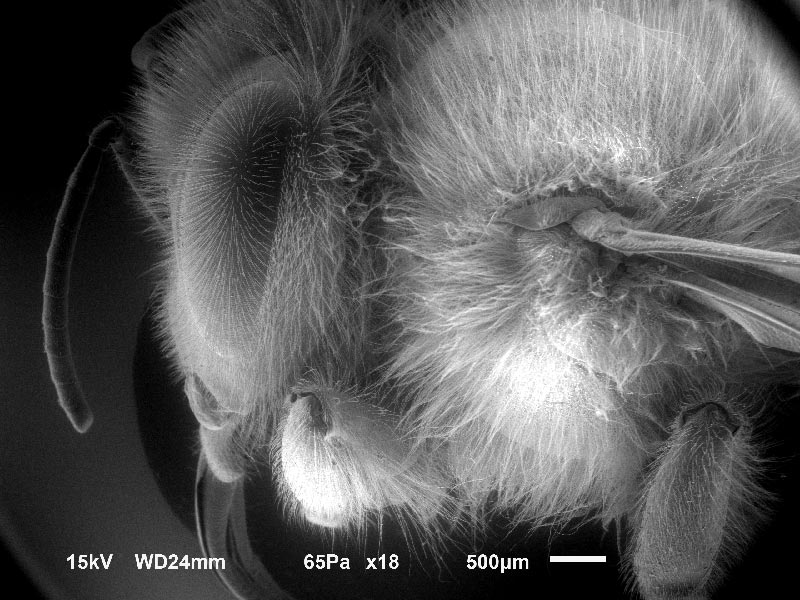
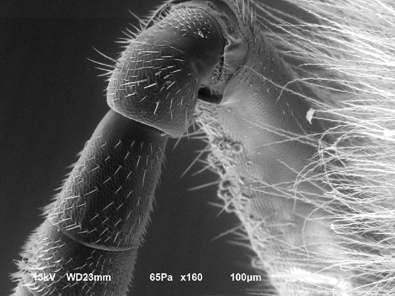
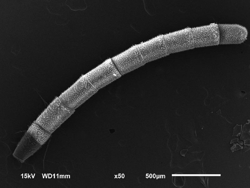
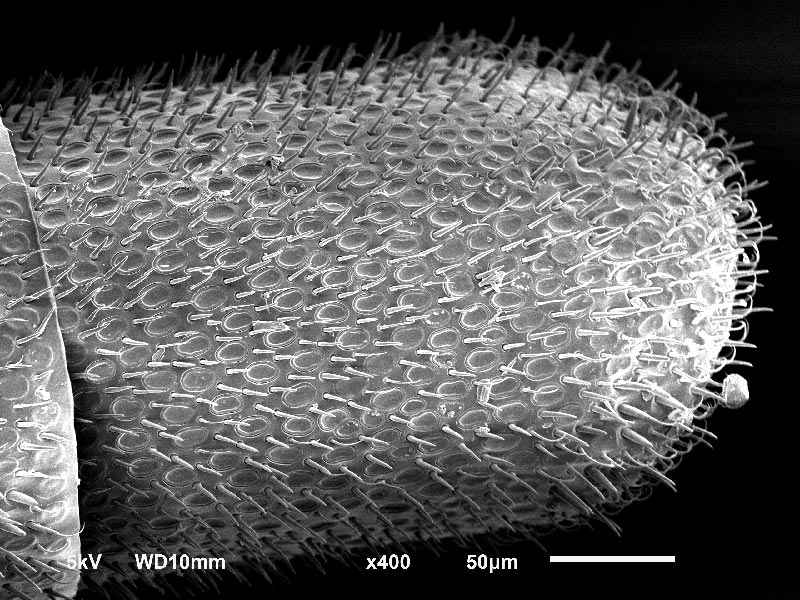
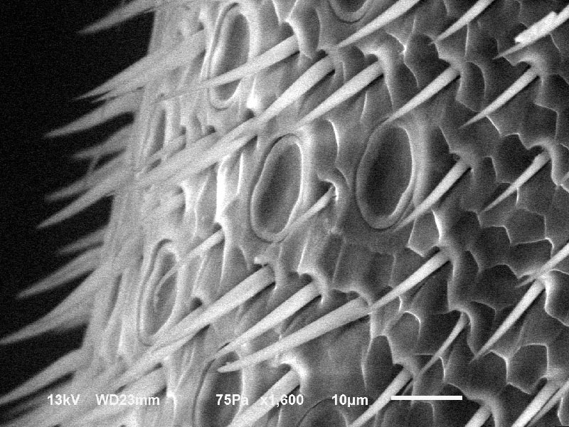
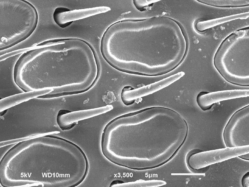
Skip to content

Honey Bee

Honey Bee

Honey Bee

Price varies by image license. 

Pictured is a honey bee in the stereo light microscope magnified 7X. Additional images include an antennae & stinger from the bee under both a stereo light microscope and an electron microscope.

Product Description

Honey Bee – Antenna

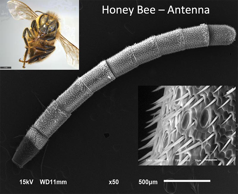
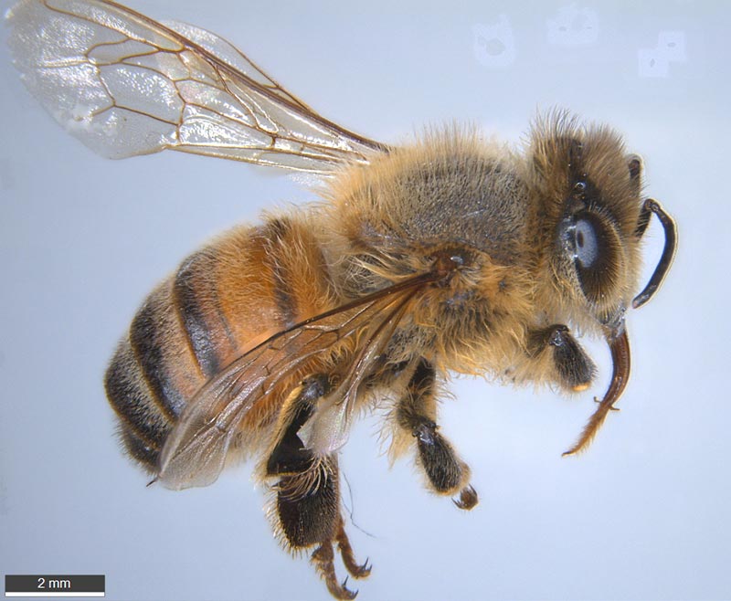
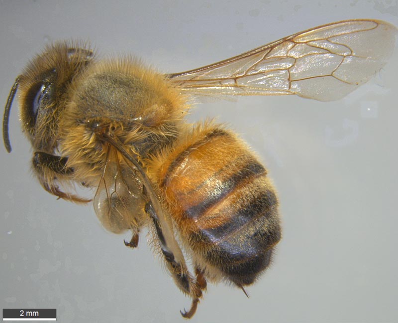
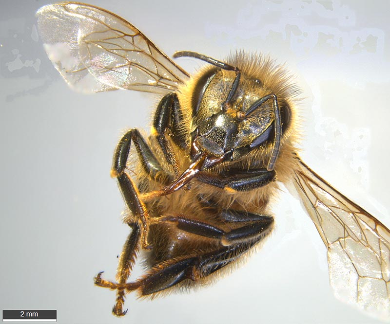
Pictured is a honey bee in the stereo light microscope magnified 7X. An antenna from the bee is put into the electron microscope magnified 50X in the center. We can see that the antennae has different electrical charges. The dark gray area at the base is more negatively charged and the tip is slightly negatively charged compared to the rest of the antennae. The surface of the antenna is magnified 1600X in the inset picture. It turns out that the ground is positively charged and flowers are negatively charged. When the bee flies through the air it becomes positively charged by the air particles hitting it. As it starts to land the negative pollen is attracted to it and it changes the field on the flower to be more positive. This field change signals other bees that the flower has been visited and the nectar supply may be low. So, the next bee that comes by will not waste his energy stopping at the flower. The antennas have many other sensing functions They include sensing magnetic fields, vibrations, flying speed, taste, scent, tactile information, photo receptors, humidity, carbon dioxide, shape and gravity.
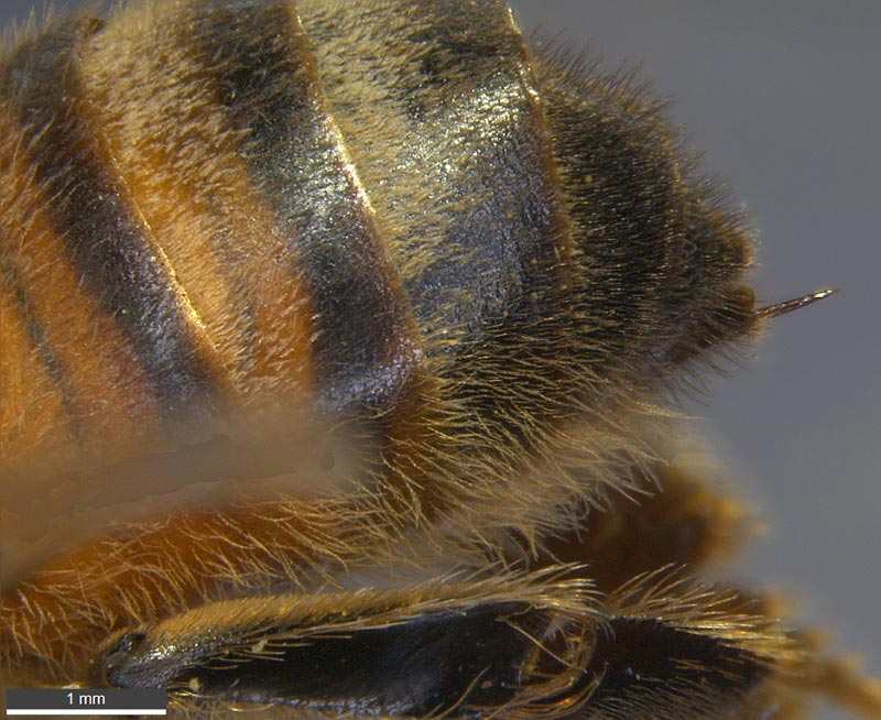
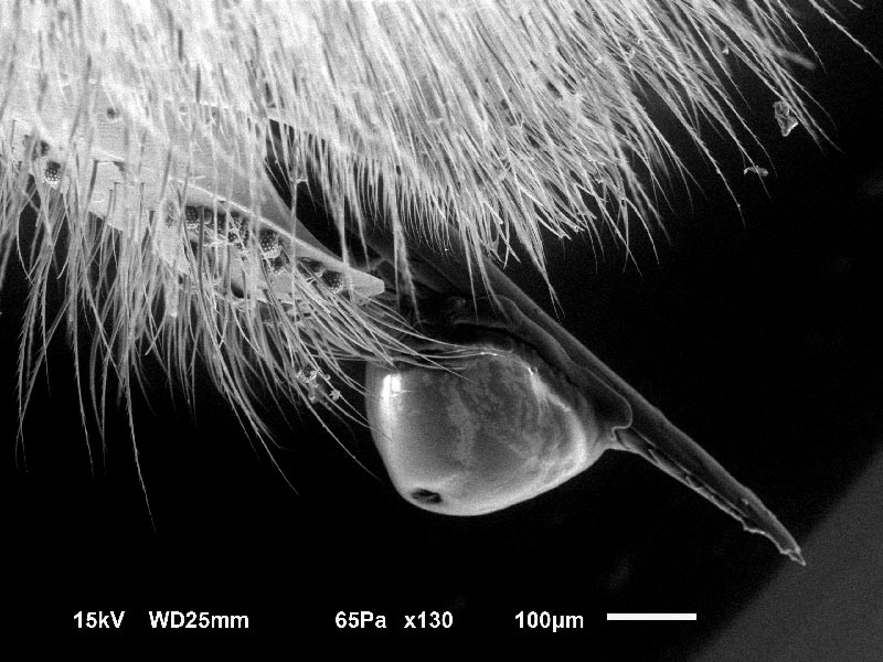
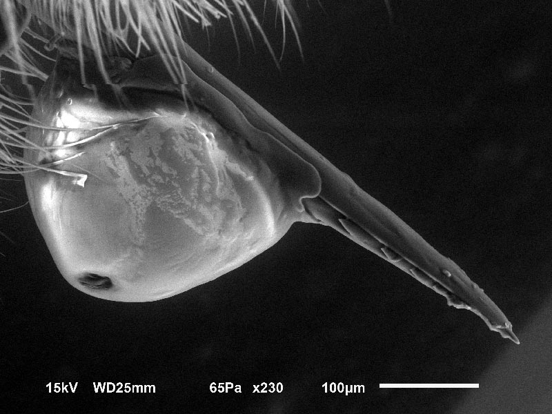
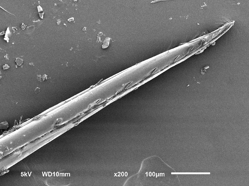
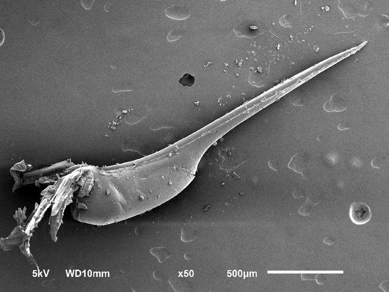
Honey Bee – Stinger

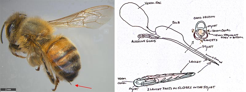
The honeybee has an effective weapon against predators. When a worker bee feels a deadly treat to itself or its nest, it stings. The honeybee stinger is hollow and pointed similar to a hypodermic needle. However, it has a 2-part lancet with rows of sharp barbs that slide on a stylet base. The barbed portion is only 0.5 mm long but goes much farther that that when jammed into the skin of a predator. It is difficult to remove and is torn from the honeybee. The bee venom is pumped through the 2 sides and bottom of the lancet quickly flooding the area to maximize the stinging impact. A pheromone is also released that warns other bees.

PURCHASE 5 OR MORE IMAGES AND GET 20% OFF YOUR ENTIRE ORDER!

Please contact us for custom images.

The image store is a collection of organisms that have been examined under a stereo light microscope (LM) and or scanning electron microscope (SEM). Each group of organisms has a short description and a longer more detailed description or story about the organism.  Clicking on the product group shows the individual images.  Each series takes the observer from macro to micro or nano on a particular organism, starting with a macro photographic image(s) for perspective, micro images taken by the light microscope, and most have micro to nano scanning electron microscope images.  The SEM images will appear in black and white as a beam of electrons is used to illuminate the specimen rather than light.  A few SEM images are colorized (lotus leaf).  More information about the labeling and techniques used is below.

For the curious:

The light microscope images are labeled LM and a Z is included if it is a vertical composite of images effectively extending the depth of field or EDF of the microscope.

SEM images are labeled by the type of detector use:

SE (secondary electron)

LSE (Low vacuum secondary electron)

BES (backscattered electron shadow mode)

BEC (backscattered electron compositional mode)

The SEM instrument works by producing a beam of electrons under a vacuum that interacts with the sample surface and subsurface producing different signals, as shown in the diagram at right.  Secondary electrons, backscattered electrons and x-rays are detected using different instrument modes.  In addition to morphological information to produce an image the SEM can determine elemental composition by energy dispersive x-ray spectroscopy (EDS).

Personal/Educational License:
For personal or educational use only.  Images cannot be given or resold to others.

Commercial License:
For Profit or Non-profit business use within the organization, in presentations and publications with image credit.  No resale of images.

An image license does not grant exclusive rights to an image.  Lichen Labs retains the copywrite to all images.

See Full Terms and Conditions