Product Description
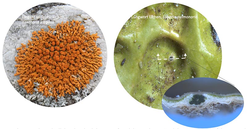
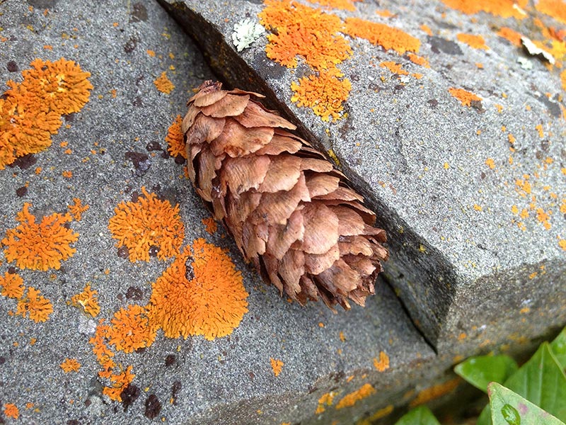
An everyday organism, the lichen inspired the name for Lichen Labs LLC. Lichens cover 8% of the terrestrial surface of the earth on rocks, trees, the forest floor, and even on this tombstone in Virginia, Minnesota (above Left). It was used as a model for the company logo. They have anchors but no roots and get their nourishment through the air. Lichens can also come back to life after years, sometimes decades, without water
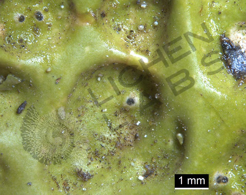
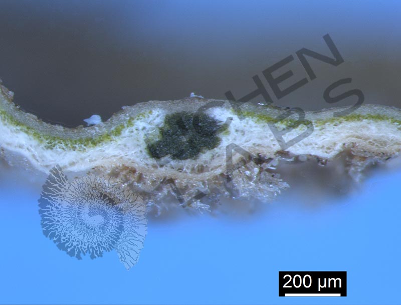
Lichens are really fungi and algae or cyanobacteria living together in the same body. The fungus provides the home, and the algae and or cyanobacteria make the food. A slice was taken of this green lungwort lichen, from a tree on Vancouver Is. Canada, to Look inside (above Right). The algae are in the green layer under the protective waxy outer layer. The very dark green dot is cyanobacteria, another organism that does photosynthesis for food.
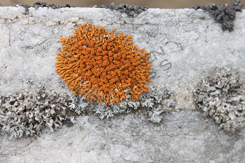
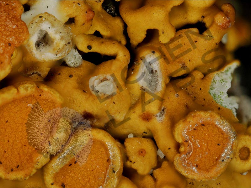
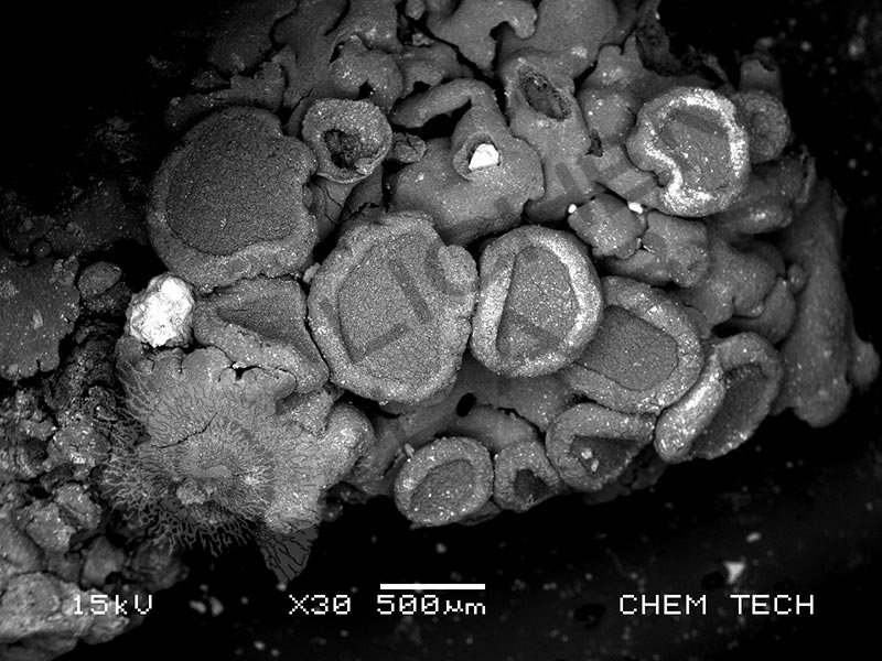
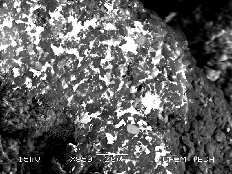
 After looking under a microscope at an orange lichen growing on a twig in the mountains of Montana, shiny areas were noticed around a rim of an apothecia structure.
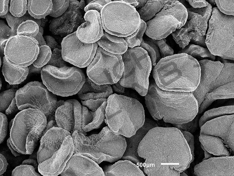
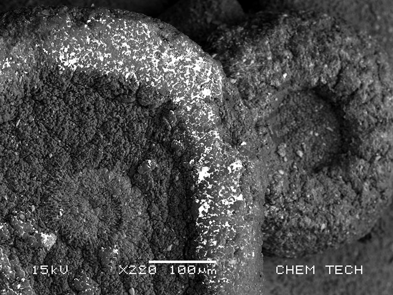
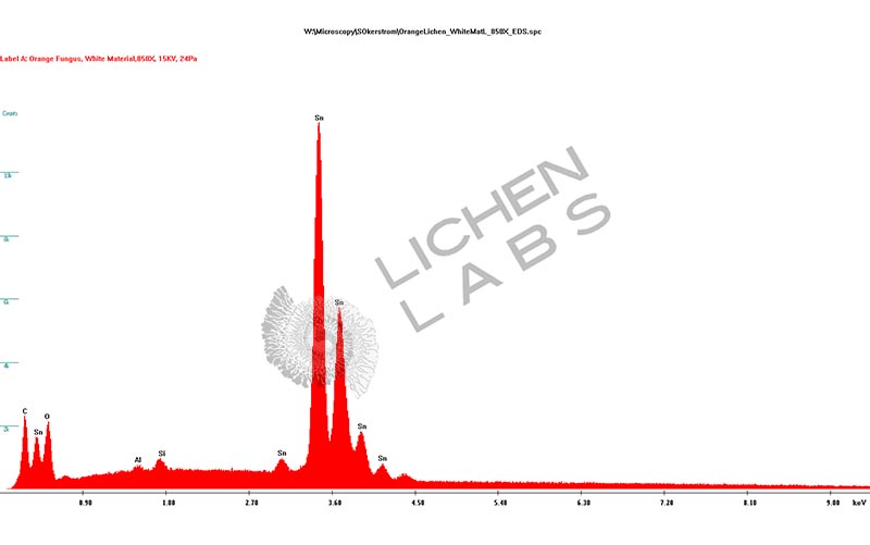
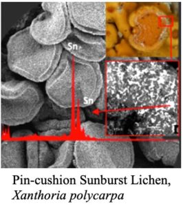
After further study in an electron microscope, that shiny area of the lichen was found to be almost entirely made of tin (Sn)! It turns out lichen are also miners and can absorb and concentrate minerals from the air. Lichens are a symbiotic relationship between fungus and algae, much like the collaborative nature of Lichen Labs. Some lichens can live for 4,000 years. Now that’s a long-term relationship!
References:
Field guide: “Lichens of the North Woods” by Joe Walewski, 2007
Bible: “Lichens of North America” by Irwin M. Brodo, Sylvia Duran Sharnoff and Stephen Sharnoff, 2001
PURCHASE 5 OR MORE IMAGES AND GET 20% OFF YOUR ENTIRE ORDER!
Please contact us for custom images.
The image store is a collection of organisms that have been examined under a stereo light microscope (LM) and or scanning electron microscope (SEM). Each group of organisms has a short description and a longer more detailed description or story about the organism. Clicking on the product group shows the individual images. Each series takes the observer from macro to micro or nano on a particular organism, starting with a macro photographic image(s) for perspective, micro images taken by the light microscope, and most have micro to nano scanning electron microscope images. The SEM images will appear in black and white as a beam of electrons is used to illuminate the specimen rather than light. A few SEM images are colorized (lotus leaf). More information about the labeling and techniques used is below.
For the curious:
The light microscope images are labeled LM and a Z is included if it is a vertical composite of images effectively extending the depth of field or EDF of the microscope.
SEM images are labeled by the type of detector use:
SE (secondary electron)
LSE (Low vacuum secondary electron)
BES (backscattered electron shadow mode)
BEC (backscattered electron compositional mode)
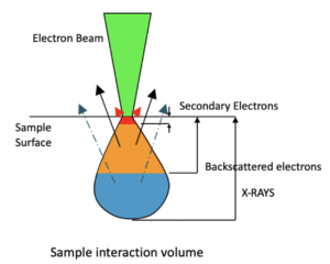
The SEM instrument works by producing a beam of electrons under a vacuum that interacts with the sample surface and subsurface producing different signals, as shown in the diagram at right. Secondary electrons, backscattered electrons and x-rays are detected using different instrument modes. In addition to morphological information to produce an image the SEM can determine elemental composition by energy dispersive x-ray spectroscopy (EDS).