Product Description
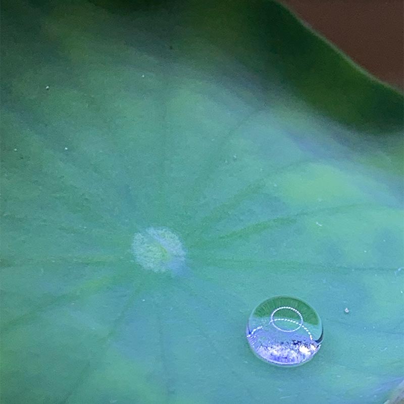
This lotus has self-cleaning leaves. The leaves appear to be very smooth, but they actually have a nano-texture of waxy pillars with air between them. When a water droplet hits the leaf, if there’s even the slightest tilt, it will roll off the surface and take dirt particles with it. This happens because dirt sticks better to the water droplets then the lotus leaf surface. Below is a slow-motion video of droplets rolling off a Lotus leaf. It is an example of super-hydrophobicity (a surface that repels water).
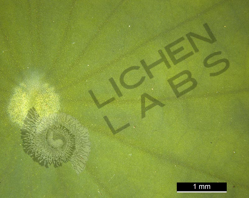
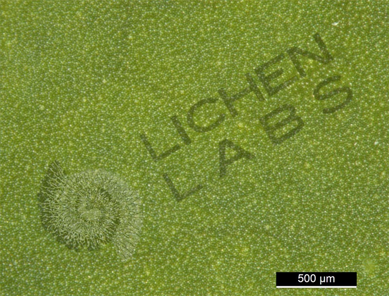
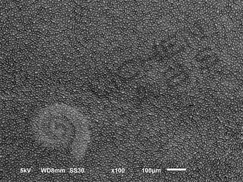
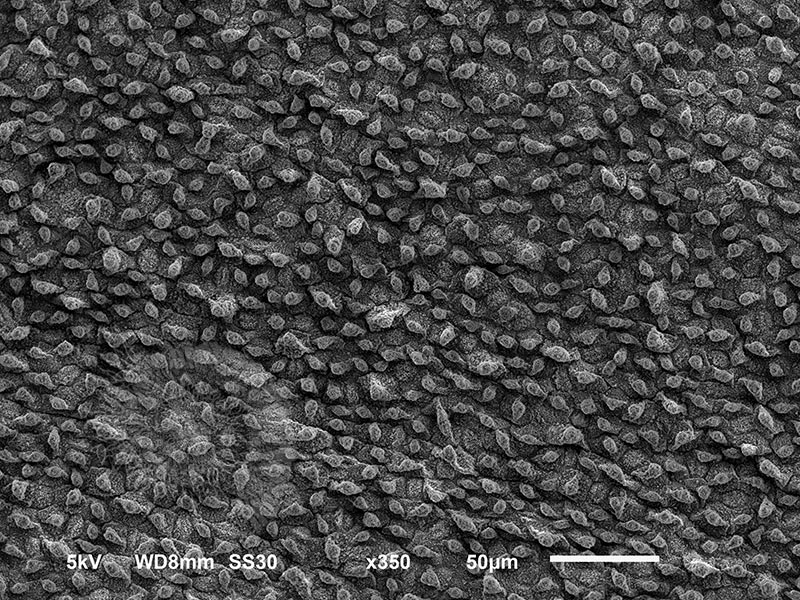
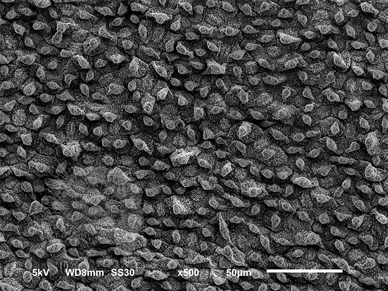
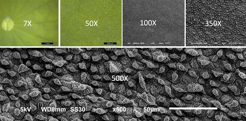
The collection of lotus leaf images below shows the surface of the leaf magnified beginning under a Leica stereo light microscope at 7X(times) and 50X then the scanning electron microscope (SEM) at 100X, 350X, 500X. The magnification corresponds to the settings on the microscope when the pictures were taken. As you can see the bottom image is magnified much more relative to the other images. To keep the “scale” of the image the same no matter how much the image is enlarged, microscopists put a scale bar on each image. It is the white bar with the length of the bar next to it. For the 500X image, the bar is 50µm or microns long. 50 µm is 50 millionths of a meter (0.000050 m) equivalent to 0.002” or the width of an average size hair.
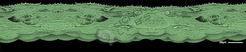
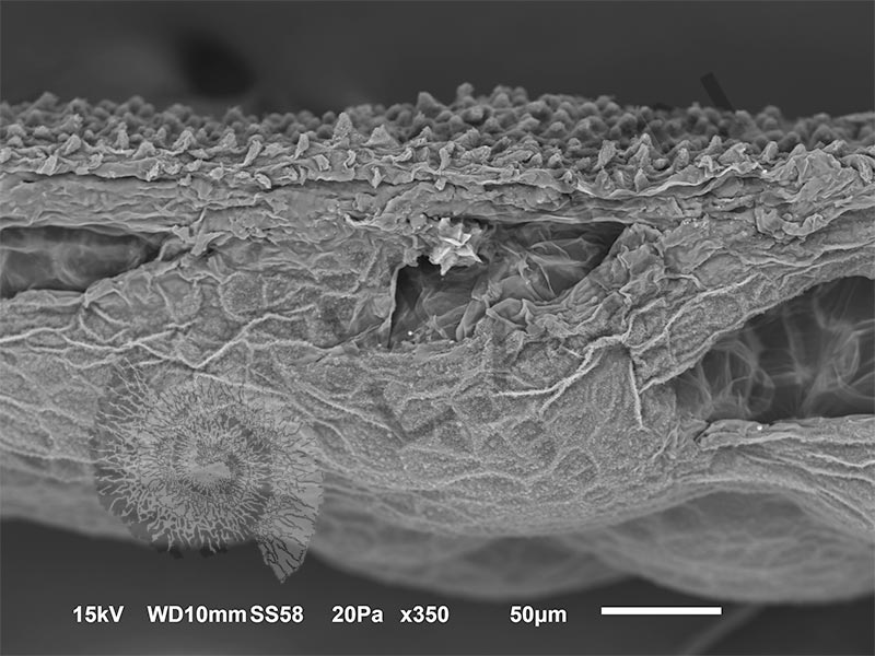
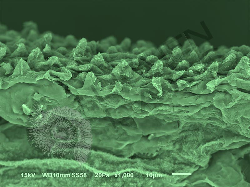
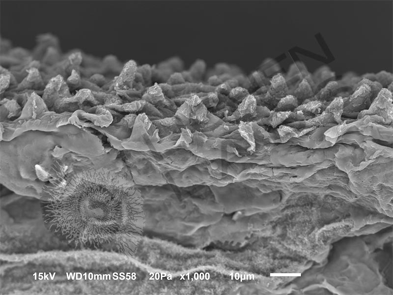
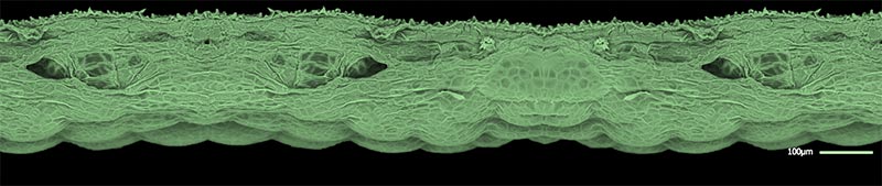
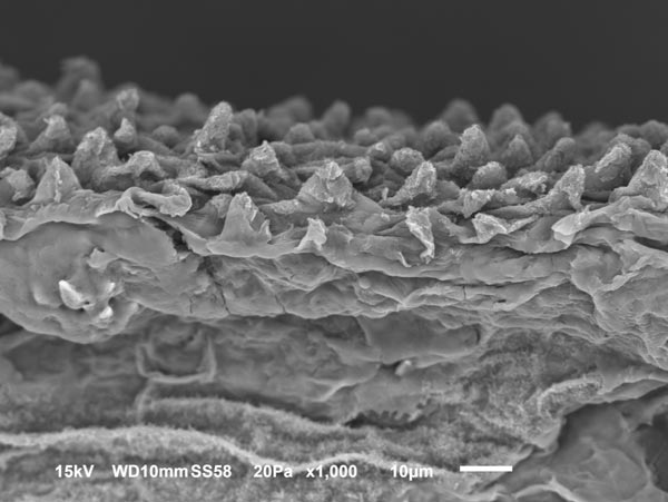
The lotus leaf was also cross-sectioned and examined in the SEM to see the side view and measure the height of the pillars. Below is a colorized composite SEM image of the leaf cross-section with a magnified view at the bottom.
Super-hydrophobicity using a micro or nanostructure is another pattern in nature used by some spiders, insects, and other plants.
PURCHASE 5 OR MORE IMAGES AND GET 20% OFF YOUR ENTIRE ORDER!
Please contact us for custom images.
The image store is a collection of organisms that have been examined under a stereo light microscope (LM) and or scanning electron microscope (SEM). Each group of organisms has a short description and a longer more detailed description or story about the organism. Clicking on the product group shows the individual images. Each series takes the observer from macro to micro or nano on a particular organism, starting with a macro photographic image(s) for perspective, micro images taken by the light microscope, and most have micro to nano scanning electron microscope images. The SEM images will appear in black and white as a beam of electrons is used to illuminate the specimen rather than light. A few SEM images are colorized (lotus leaf). More information about the labeling and techniques used is below.
For the curious:
The light microscope images are labeled LM and a Z is included if it is a vertical composite of images effectively extending the depth of field or EDF of the microscope.
SEM images are labeled by the type of detector use:
SE (secondary electron)
LSE (Low vacuum secondary electron)
BES (backscattered electron shadow mode)
BEC (backscattered electron compositional mode)
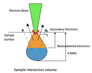
The SEM instrument works by producing a beam of electrons under a vacuum that interacts with the sample surface and subsurface producing different signals, as shown in the diagram at right. Secondary electrons, backscattered electrons and x-rays are detected using different instrument modes. In addition to morphological information to produce an image the SEM can determine elemental composition by energy dispersive x-ray spectroscopy (EDS).