Product Description
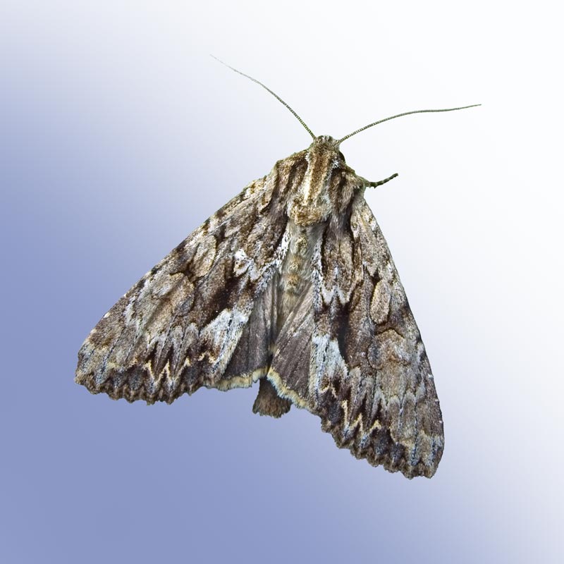
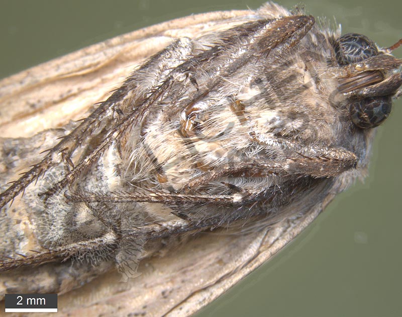
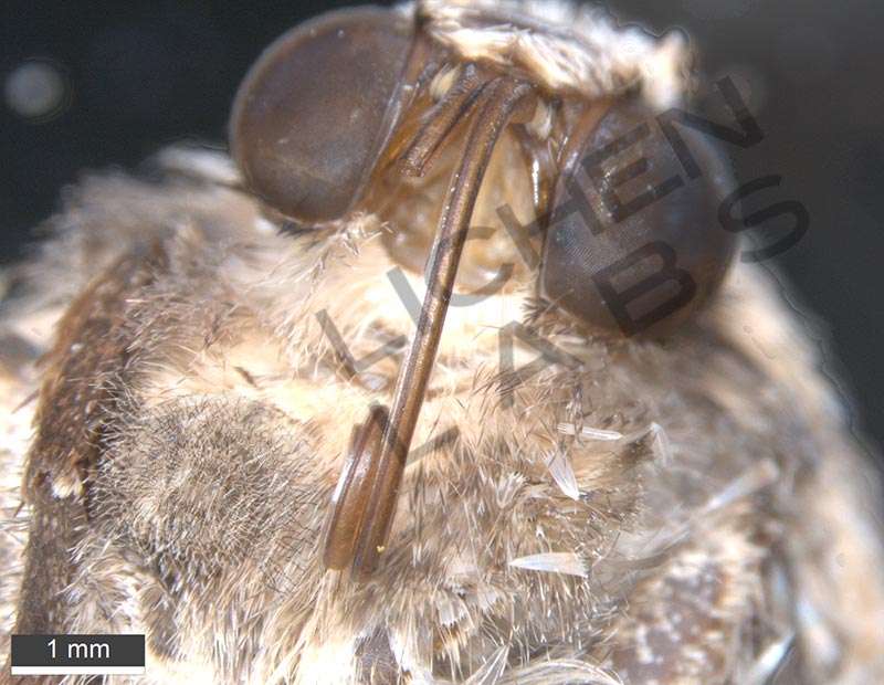
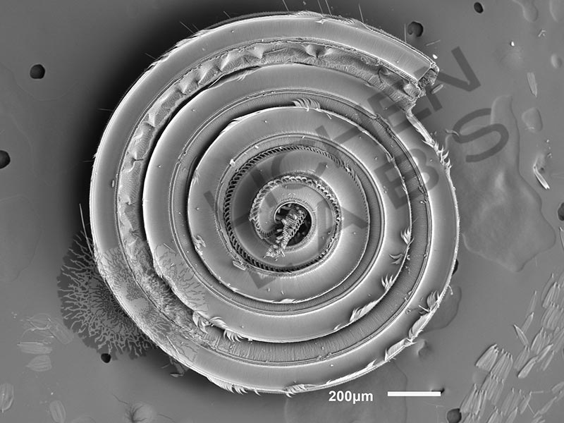
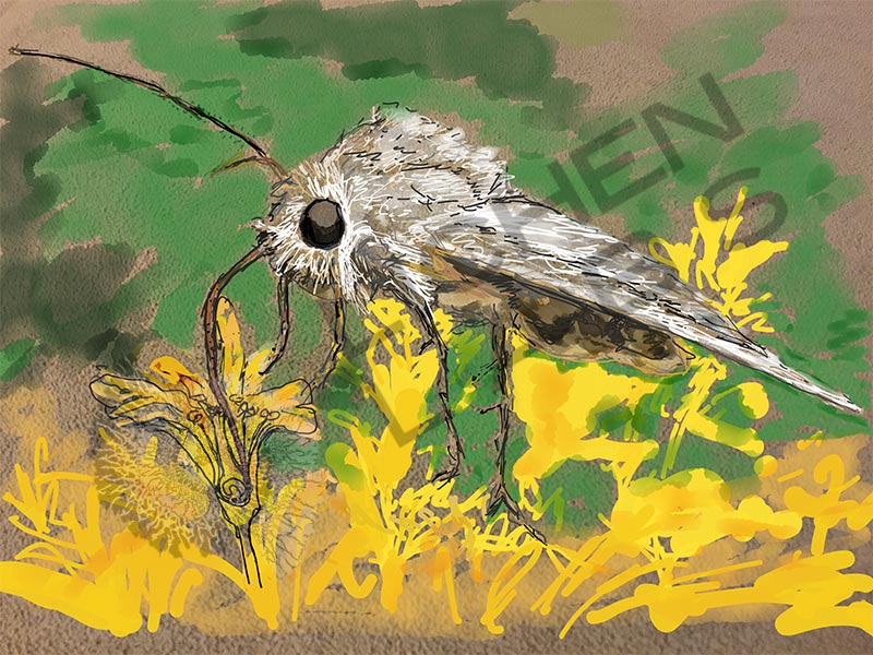
The noctuid moth has adapted the size and shape of structures needed to feed from flowers in their habitat. Sweet sugary nectar is made by the flowers to attract insects to assist in the pollination process. This 24 mm long moth has a 10 mm long extended proboscis for sucking up nectar.
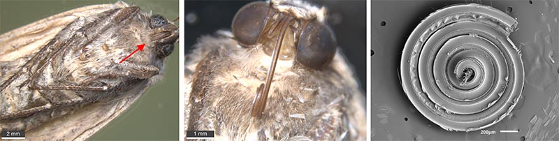

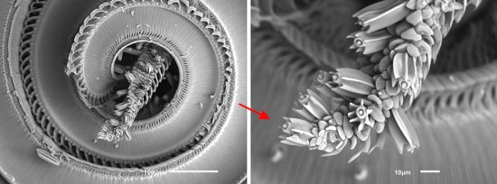

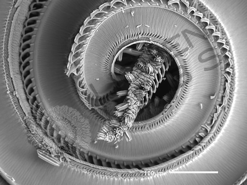
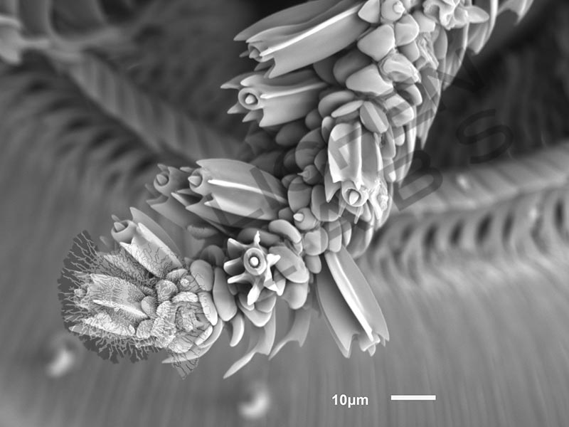
A sucking tube is formed by linking together in a series of hooks and plates between 2 sets of maxillary muscles allowing the long proboscis to coil up under the head when not in use. The tongue like tips has several types of sensing organs called sensilla that enable the moth to locate and taste the nectar. The moth rolls out the proboscis using the maxillary muscles when it is ready to feed and probes into the flower brushing against pollen containing reproductive organs. The quality of the nectar is checked with the probosci’s tip sensilla which consists of a sensory cone in the center of the pore at the end with 6 or 7 longitudinal flat fins encircling it that appears to protect it when it is extended into the base of the flower. The nectar is tasted then sucked in through slits in the tube by expansion and contraction of a sack in its head.
PURCHASE 5 OR MORE IMAGES AND GET 20% OFF YOUR ENTIRE ORDER!
Please contact us for custom images.
The image store is a collection of organisms that have been examined under a stereo light microscope (LM) and or scanning electron microscope (SEM). Each group of organisms has a short description and a longer more detailed description or story about the organism. Clicking on the product group shows the individual images. Each series takes the observer from macro to micro or nano on a particular organism, starting with a macro photographic image(s) for perspective, micro images taken by the light microscope, and most have micro to nano scanning electron microscope images. The SEM images will appear in black and white as a beam of electrons is used to illuminate the specimen rather than light. A few SEM images are colorized (lotus leaf). More information about the labeling and techniques used is below.
For the curious:
The light microscope images are labeled LM and a Z is included if it is a vertical composite of images effectively extending the depth of field or EDF of the microscope.
SEM images are labeled by the type of detector use:
SE (secondary electron)
LSE (Low vacuum secondary electron)
BES (backscattered electron shadow mode)
BEC (backscattered electron compositional mode)
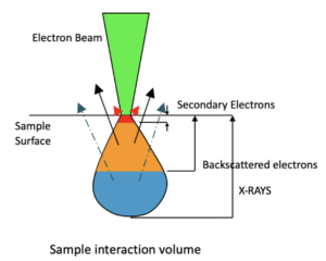
The SEM instrument works by producing a beam of electrons under a vacuum that interacts with the sample surface and subsurface producing different signals, as shown in the diagram at right. Secondary electrons, backscattered electrons and x-rays are detected using different instrument modes. In addition to morphological information to produce an image the SEM can determine elemental composition by energy dispersive x-ray spectroscopy (EDS).