Product Description
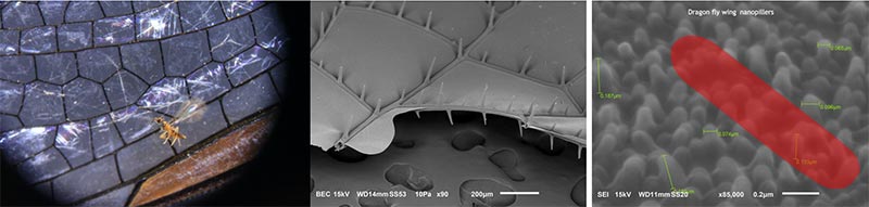
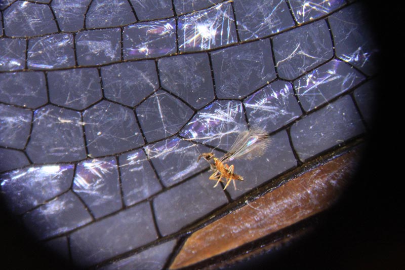
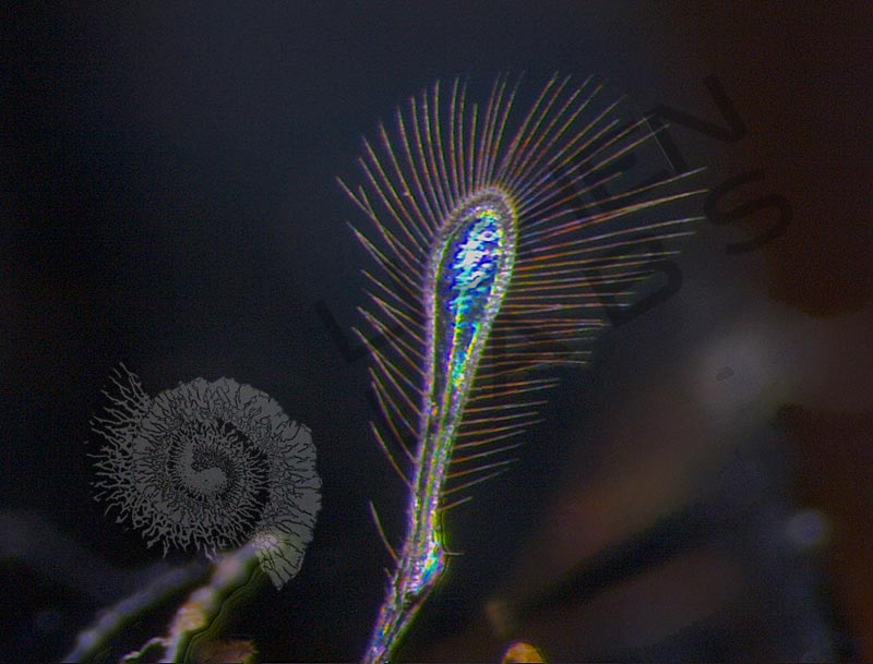
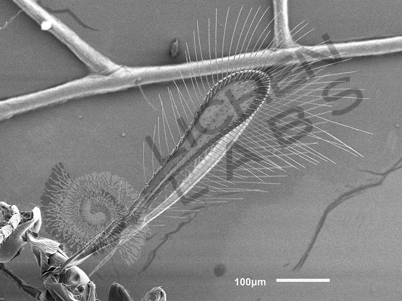
A biomimicry colleague from Mexico City and I did an “expedition” to the North Shore of Lake Superior. We were collecting small samples and insects to examine their structural adaptations under a microscope. One specimen was an eastern blue darner dragon fly that happened to be stuck to the bumper of my Mini Cooper. Back in the lab in Minneapolis, I looked at the dragon fly wing under a microscope. There was a glint of gold near the edge. Bringing it into focus revealed a tiny insect a fraction of a millimeter long with fairy-like iridescent feathery wings (above left).
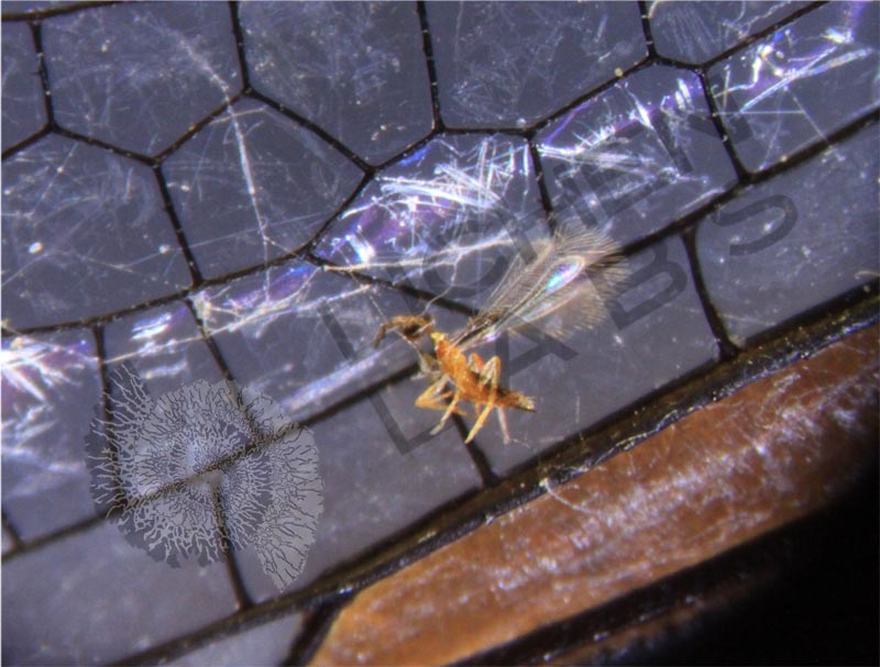
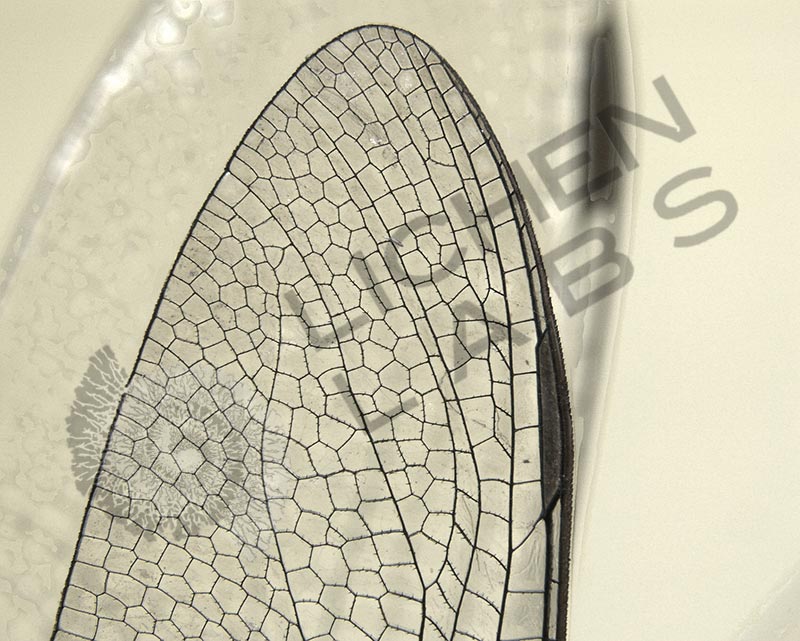
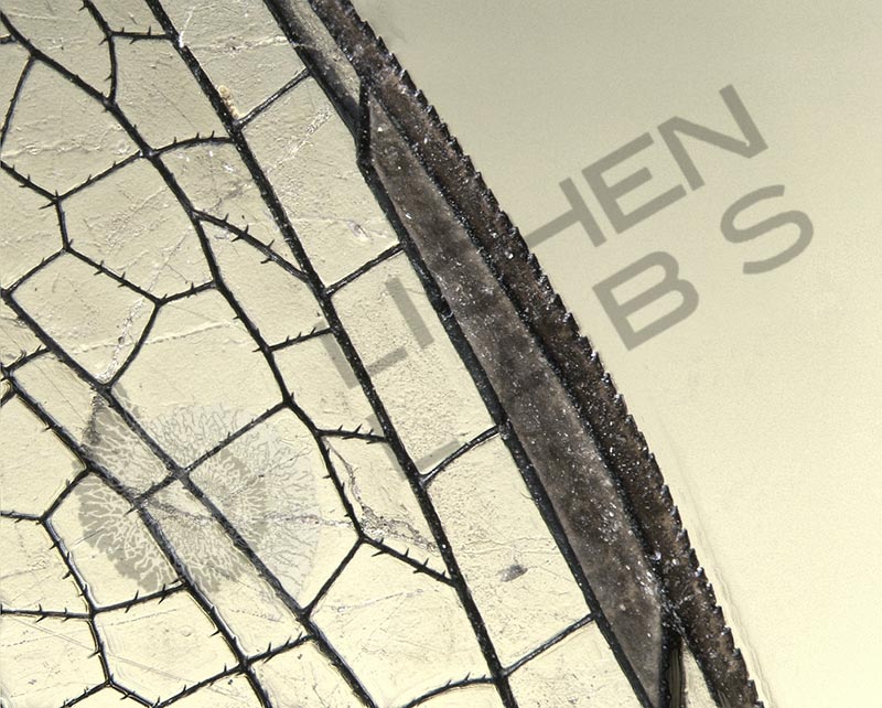
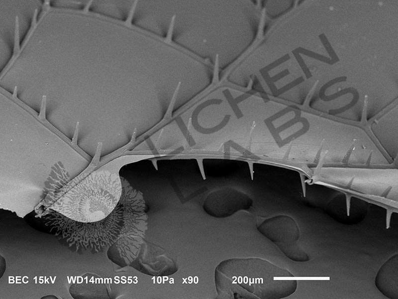
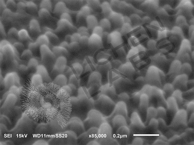
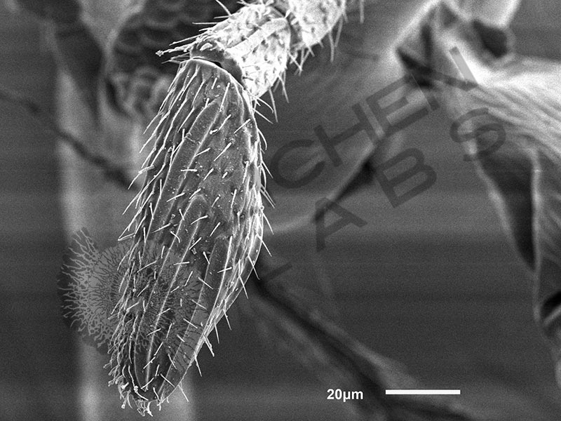
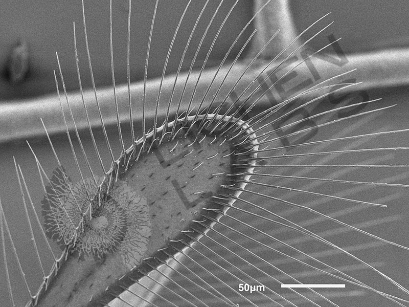
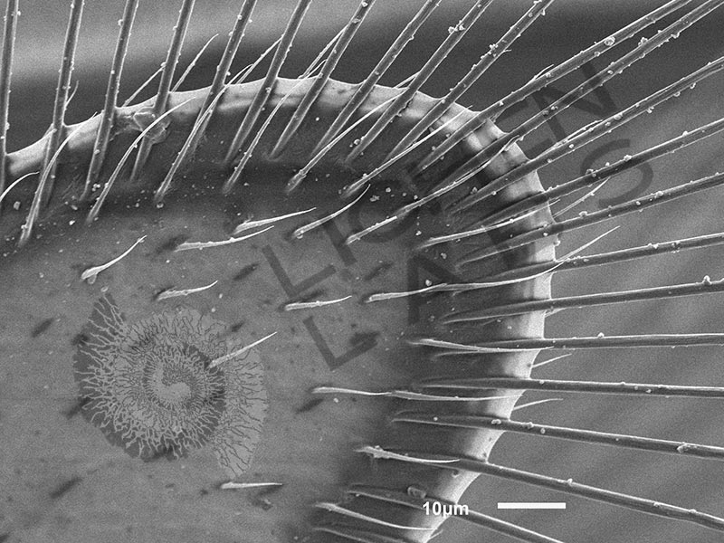
After some searching, it turned out to be a “fairy fly” parasitoid wasp in the Mymaridae family. They lay their eggs in the eggs of their insect host, which are often underwater. Their wing beat is similar to a hummingbird in speed but serve a multi-functional purpose. The fairy fly’s wings can be used as paddles underwater. The image above left is the fairy fly on the dragon fly wing. The fairy fly evidently was in the air and caught on the spikes on the dragon fly wing structure (above center). On the nanoscale, dragon fly wing surfaces have nanopillars (the texture in the image above right) that are anti-bacterial. Bacteria that land on the wing stick on the nanopillers and are ruptured. A Bacillus subtilis bacteria rod is drawn to scale and superimposed on the nanopillers on the wing surface to compare size.
PURCHASE 5 OR MORE IMAGES AND GET 20% OFF YOUR ENTIRE ORDER!
Please contact us for custom images.
The image store is a collection of organisms that have been examined under a stereo light microscope (LM) and or scanning electron microscope (SEM). Each group of organisms has a short description and a longer more detailed description or story about the organism. Clicking on the product group shows the individual images. Each series takes the observer from macro to micro or nano on a particular organism, starting with a macro photographic image(s) for perspective, micro images taken by the light microscope, and most have micro to nano scanning electron microscope images. The SEM images will appear in black and white as a beam of electrons is used to illuminate the specimen rather than light. A few SEM images are colorized (lotus leaf). More information about the labeling and techniques used is below.
For the curious:
The light microscope images are labeled LM and a Z is included if it is a vertical composite of images effectively extending the depth of field or EDF of the microscope.
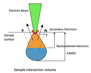
SEM images are labeled by the type of detector use:
SE (secondary electron)
LSE (Low vacuum secondary electron)
BES (backscattered electron shadow mode)
BEC (backscattered electron compositional mode)
The SEM instrument works by producing a beam of electrons under a vacuum that interacts with the sample surface and subsurface producing different signals, as shown in the diagram at right. Secondary electrons, backscattered electrons and x-rays are detected using different instrument modes. In addition to morphological information to produce an image the SEM can determine elemental composition by energy dispersive x-ray spectroscopy (EDS).