Product Description
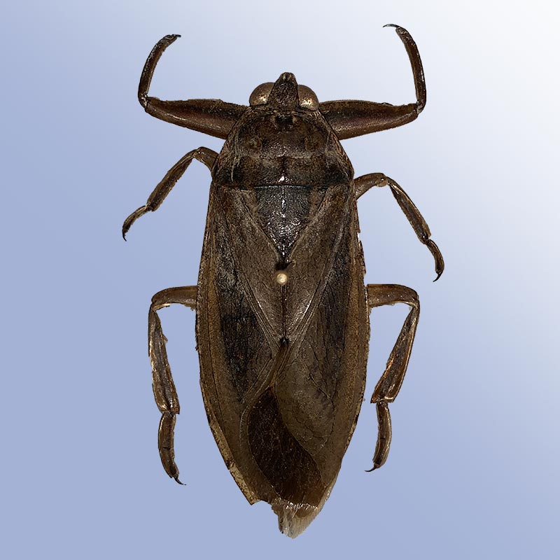
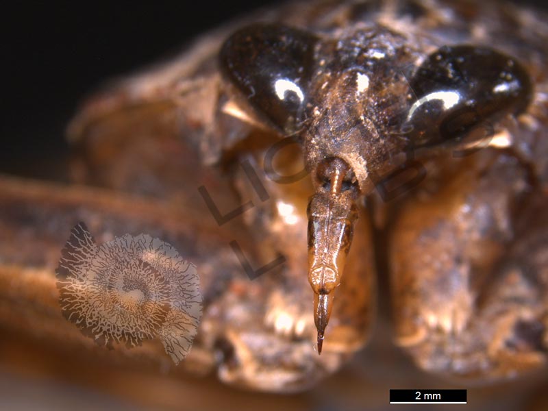
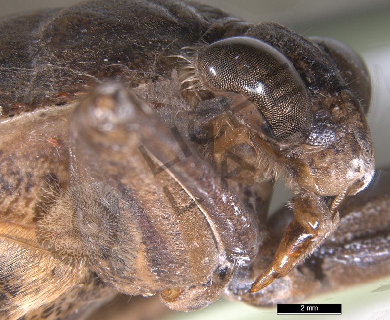
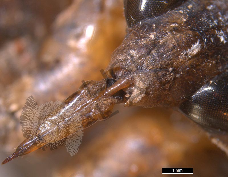
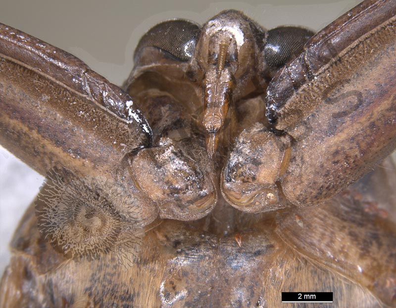
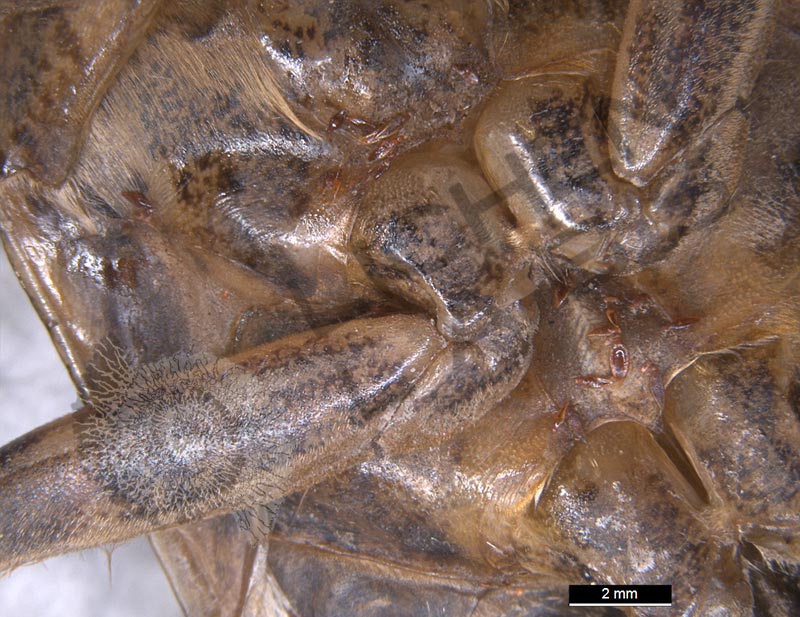
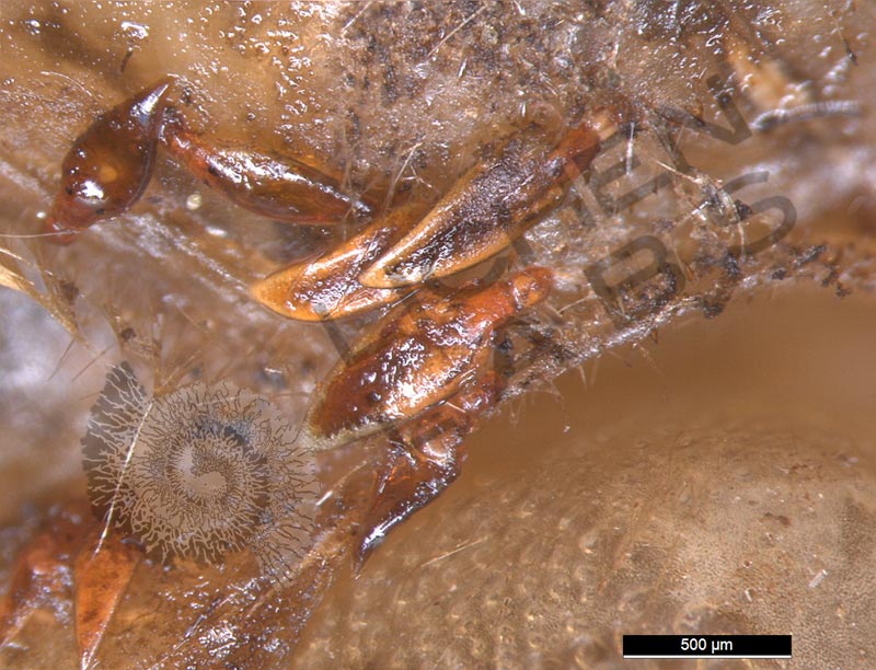
The giant water bug is true to its name – Giant and a “true bug” aquatic insect in the Belostomatidae family. They eat other insects, tadpoles, snails, small fish, and frogs. Giant water bugs have raptorial front legs made for grasping slippery prey. With a pierce from their mouth apparatus, called a rostrum, they inject toxic enzymes into their prey. After ~ 15 minutes, the prey is sufficiently digested to consume by sucking through their rostrum. It may sound unappetizing, but in southeast Asian countries, giant water bugs are a delicacy. They encounter humans most often when they fly out of their watery home at night in search of other water bodies or mates using the moon and stars as beacons to orient their flight.
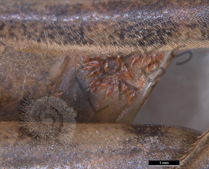
Giant water bugs get confused when encountering a light source on the ground. They fly in spirals toward the light, looking for another beacon which is impossible to see. As with moths and other insects, they will circle until they become exhausted. The female of this species lays her eggs in the shallows in aquatic vegetation, and the male keeps them moist and protects them from predators until they hatch in 1-2 weeks. This giant water bug also happened to have tiny orange aquatic mites (Hydrachnidiae) attached to its body.
http://www.naturenorth.com/summer/bug/Giant_Water_Bug.html
https://www.youtube.com/watch?v=t5nh50xMSQ4
https://en.wikipedia.org/wiki/Lethocerus_americanus
PURCHASE 5 OR MORE IMAGES AND GET 20% OFF YOUR ENTIRE ORDER!
Please contact us for custom images.
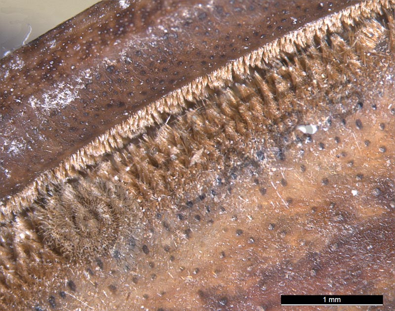
The image store is a collection of organisms that have been examined under a stereo light microscope (LM) and or scanning electron microscope (SEM). Each group of organisms has a short description and a longer more detailed description or story about the organism. Clicking on the product group shows the individual images. Each series takes the observer from macro to micro or nano on a particular organism, starting with a macro photographic image(s) for perspective, micro images taken by the light microscope, and most have micro to nano scanning electron microscope images. The SEM images will appear in black and white as a beam of electrons is used to illuminate the specimen rather than light. A few SEM images are colorized (lotus leaf). More information about the labeling and techniques used is below.
For the curious:
The light microscope images are labeled LM and a Z is included if it is a vertical composite of images effectively extending the depth of field or EDF of the microscope.
SEM images are labeled by the type of detector use:
SE (secondary electron)
LSE (Low vacuum secondary electron)
BES (backscattered electron shadow mode)
BEC (backscattered electron compositional mode)
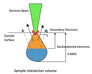
The SEM instrument works by producing a beam of electrons under a vacuum that interacts with the sample surface and subsurface producing different signals, as shown in the diagram at right. Secondary electrons, backscattered electrons and x-rays are detected using different instrument modes. In addition to morphological information to produce an image the SEM can determine elemental composition by energy dispersive x-ray spectroscopy (EDS).