Product Description
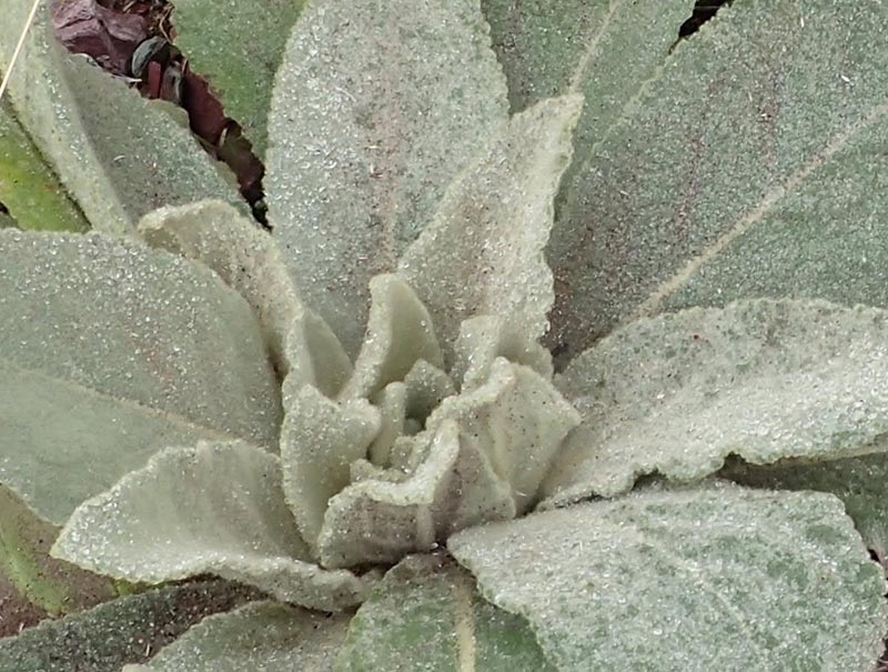
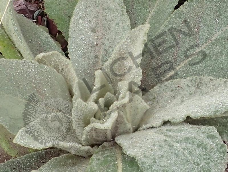
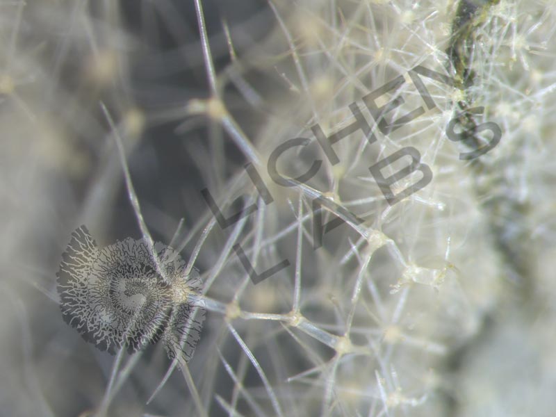
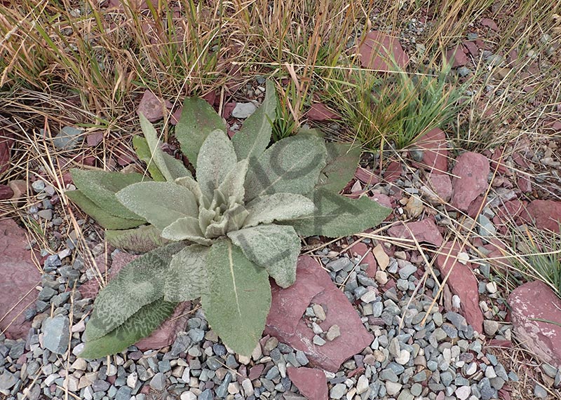
This Mullein plant (Verbascum thapsus) lives in a dry area in the mountains of Montana. This plant is also called “bunny’s ear.” Its leaves are covered in very soft hair-like appendages called trichomes. Trichomes are structures of the plant epidermis that occur on plant stems, leaves and even flowers and may be unicellular or multicellular. They vary greatly in morphology, size, and ability to secrete fluids depending on the context of their environment.
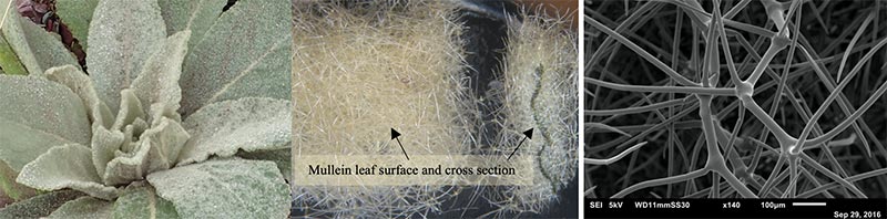
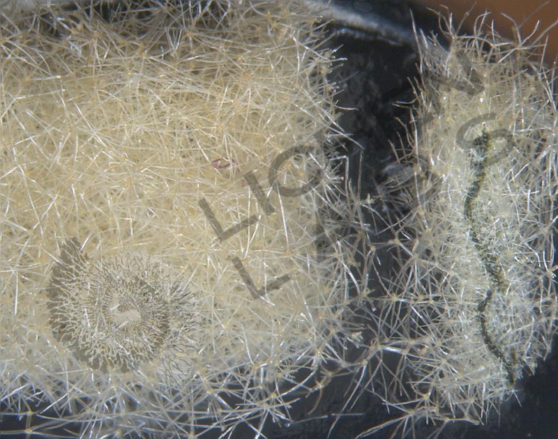
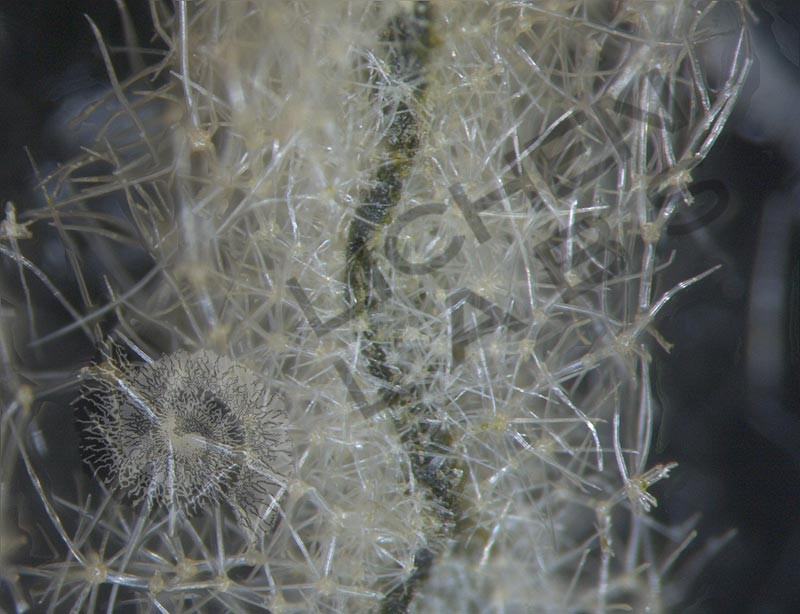
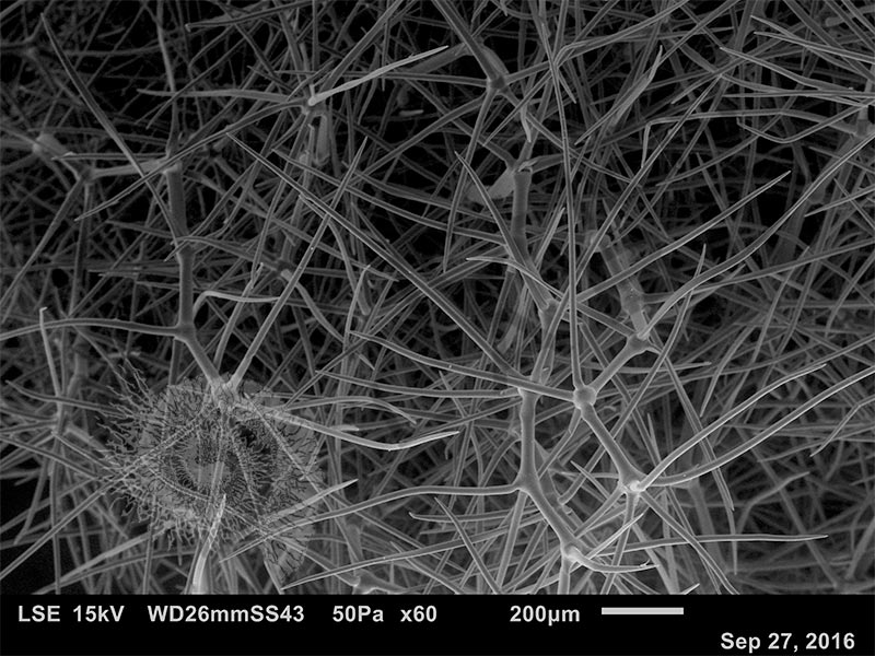
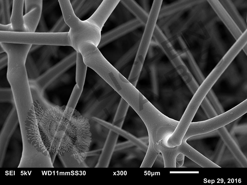
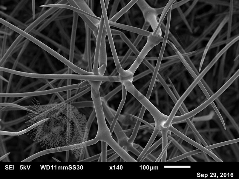
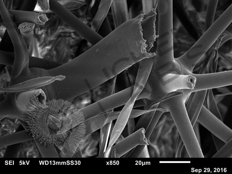
In the case of the Mullein, they adapted to protect the plant from intense solar radiation by reflecting and shading the plant leaves and preventing evaporation of moisture. The thick fuzzy mat surface (above center) also protects the leaves from being eaten by insects. Magnified in the SEM (above right), we can see that these trichomes are like layers or tiny tinker toys of branching tubes. The tubes are lightweight and insulating.
References:
http://www.biologydiscussion.com/botany/trichomes-types-development-and-functions-with-diagrams/20288
https://www.microscopemaster.com/trichomes-and-microscopy.html
PURCHASE 5 OR MORE IMAGES AND GET 20% OFF YOUR ENTIRE ORDER!
Please contact us for custom images.
The image store is a collection of organisms that have been examined under a stereo light microscope (LM) and or scanning electron microscope (SEM). Each group of organisms has a short description and a longer more detailed description or story about the organism. Clicking on the product group shows the individual images. Each series takes the observer from macro to micro or nano on a particular organism, starting with a macro photographic image(s) for perspective, micro images taken by the light microscope, and most have micro to nano scanning electron microscope images. The SEM images will appear in black and white as a beam of electrons is used to illuminate the specimen rather than light. A few SEM images are colorized (lotus leaf). More information about the labeling and techniques used is below.
For the curious:
The light microscope images are labeled LM and a Z is included if it is a vertical composite of images effectively extending the depth of field or EDF of the microscope.
SEM images are labeled by the type of detector use:
SE (secondary electron)
LSE (Low vacuum secondary electron)
BES (backscattered electron shadow mode)
BEC (backscattered electron compositional mode)
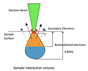
The SEM instrument works by producing a beam of electrons under a vacuum that interacts with the sample surface and subsurface producing different signals, as shown in the diagram at right. Secondary electrons, backscattered electrons and x-rays are detected using different instrument modes. In addition to morphological information to produce an image the SEM can determine elemental composition by energy dispersive x-ray spectroscopy (EDS).