

Product Description
This collection of images includes 2 different species of orb weaver spiders. It includes views of webs and web weaving spinnerets in the scanning electron microscope.
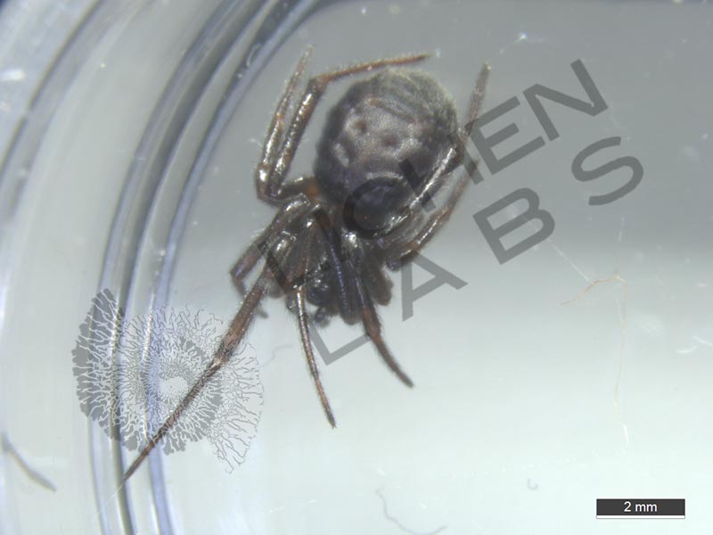
6 spotted orb weaver spiders and another species of orb weaver spider

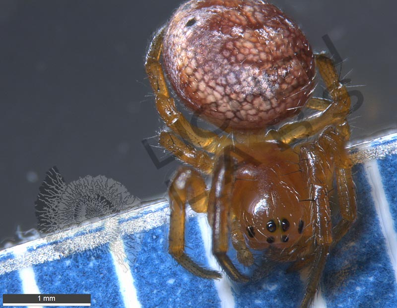
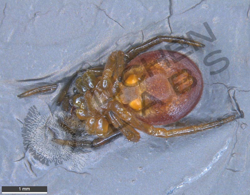
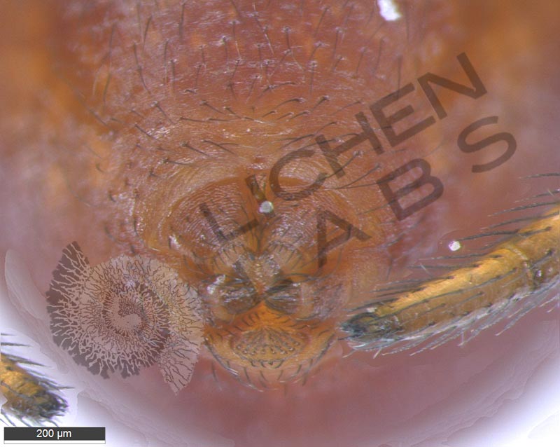
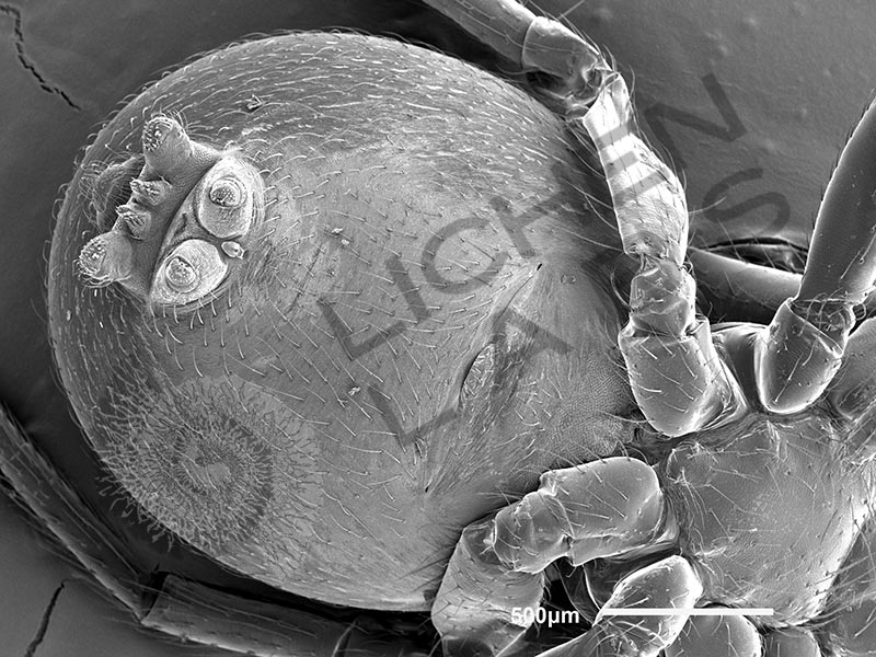
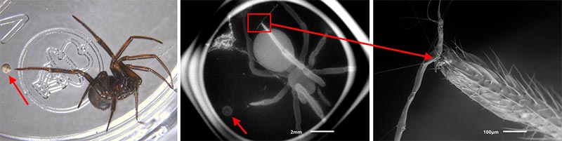
An overview image of a six spotted orb weaver spider is at left with the bottom of the spider in the center. The web spinnerets at the back of the spider are more visible in the scanning electron microscope picture at right.
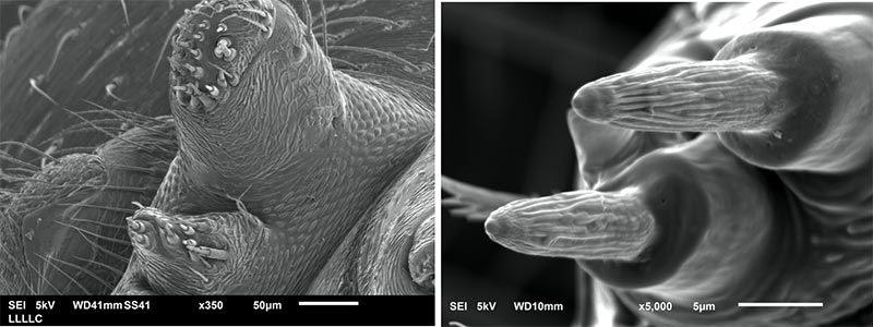
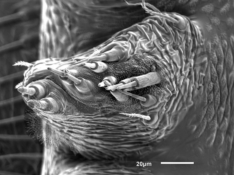
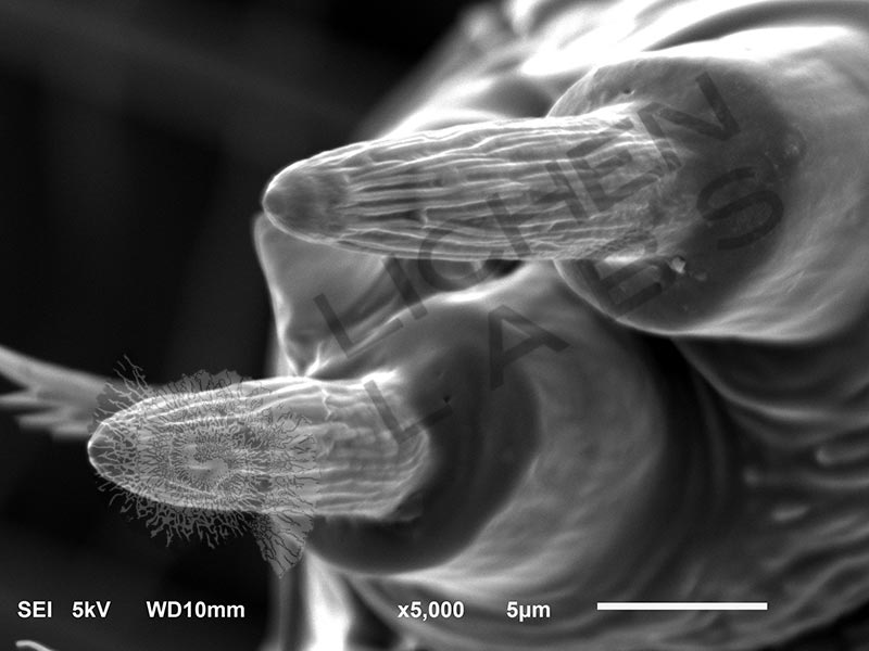
Above left is an SEM image of 2 of the spinnerets with multiple nozzles. The nozzle tips are further magnified in the center. The six spotted orb weaver spider spins small inconspicuous webs between plants near the ground to catch insects. The spider waits in the center of the web or hides at the side then rushes out to bite or wrap the insect caught in the sticky threads. In the autumn, it attaches a mass of eggs next to the web. Either the eggs overwinter, or hatch or the spiderlings overwinter. The adults die. Deep snow helps more of the young to survive and disperse in early spring.
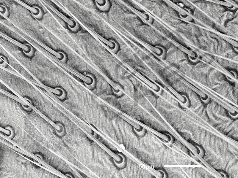
Orb weaver spiders have many silk glands in their abdomen that connect to specific spinnerets that they use to spin their webs. Orb weavers make up to 8 different types of silk, including one type of silk used to spin the egg sack in the females, another to make a sticky glue, and another for making a dragline. The dragline is a strong stiff strand of silk that forms the framework of the web or a line that the spider uses in swinging off to a new location.
The orb weaver and most spiders have 3 pairs of spinnerets on the bottom posterior end of the abdomen. Each has many spigots with a valve at the base. A chemical reaction takes place. and the liquid silk hardens as it is pulled from the spigot with the hind legs or by the weight of the spider. The spider has musculature to move the spigots independently but work together in a coordinated fashion, lifting, lowering and twisting to weave the web. The muscles also control the amount the valve opens to change the diameter of the silk. The dragline is less than 2 µm thick or 1/25th the diameter of an average human hair. The diameter depends on the size of the spider. It is constructed to hold 4 to 6 times the spider’s weight without failing. It has both high strength and elasticity. It is tougher than steel.
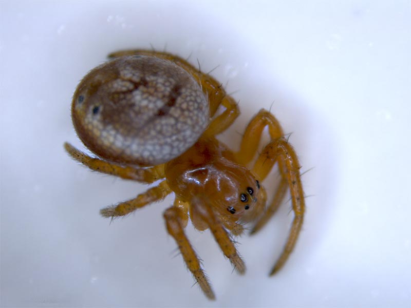
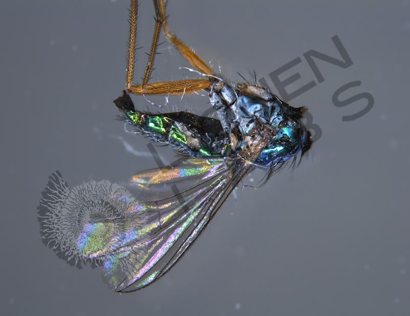
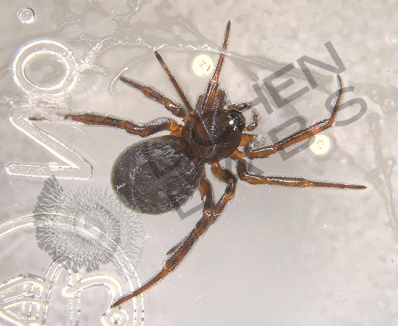
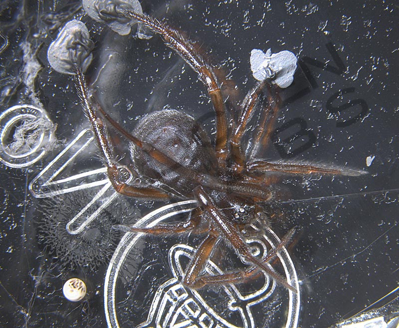
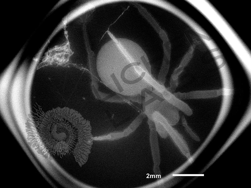
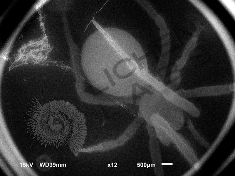
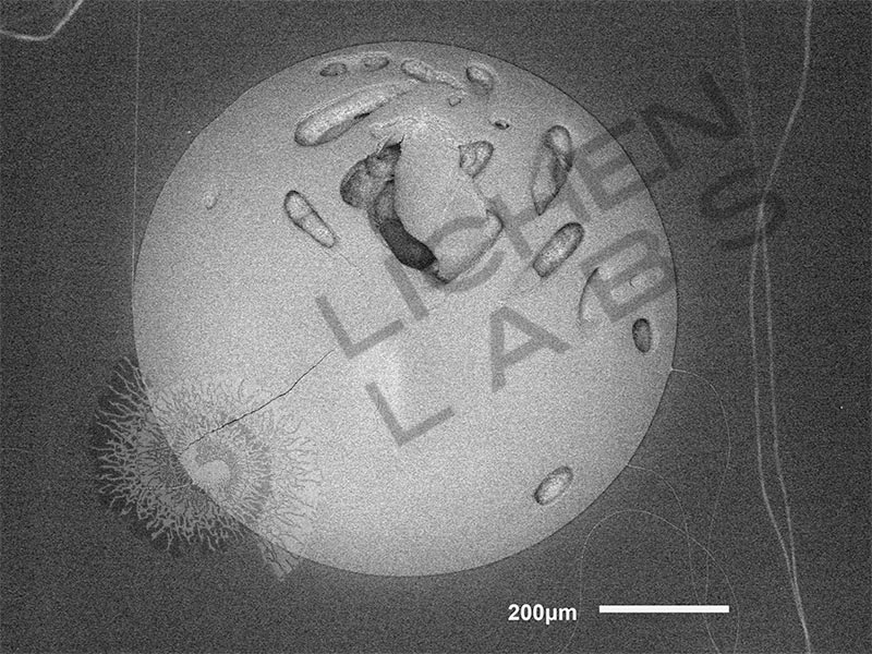
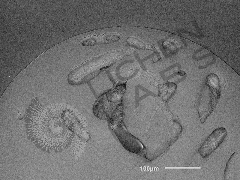
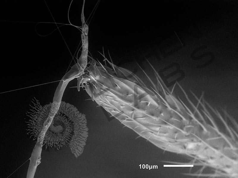
Above is a second type of orb weaver spider. It was discovered in my daughter’s bedroom and transferred to a small plastic jar where it spun a web and laid an egg. The whole jar was put in a scanning electron microscope under a vacuum – an amazing feat (below center). It was partially glued down to keep it in place. The fine web attaching its foot to the web tag line is 1 nm in diameter and is holding up the weight of the spider’s entire leg! The fine thread enables the spider to feel the slightest vibration of prey stuck on the web.
References:
“National Audubon Society Field Guide to North American Insects & Spiders” by Lorus and Margery Milne,
Copyright 1980, Thirteenth printing July 2014, Alfred A. Knopf, Publisher, New York, P.884, plate 680
“Biology of Spiders, 3rd Edition” by Rainer Foelix
MIL EAN/ISBN: 9781282917750, p.140-141,
https://en.wikipedia.org/wiki/Spider_silk