Product Description
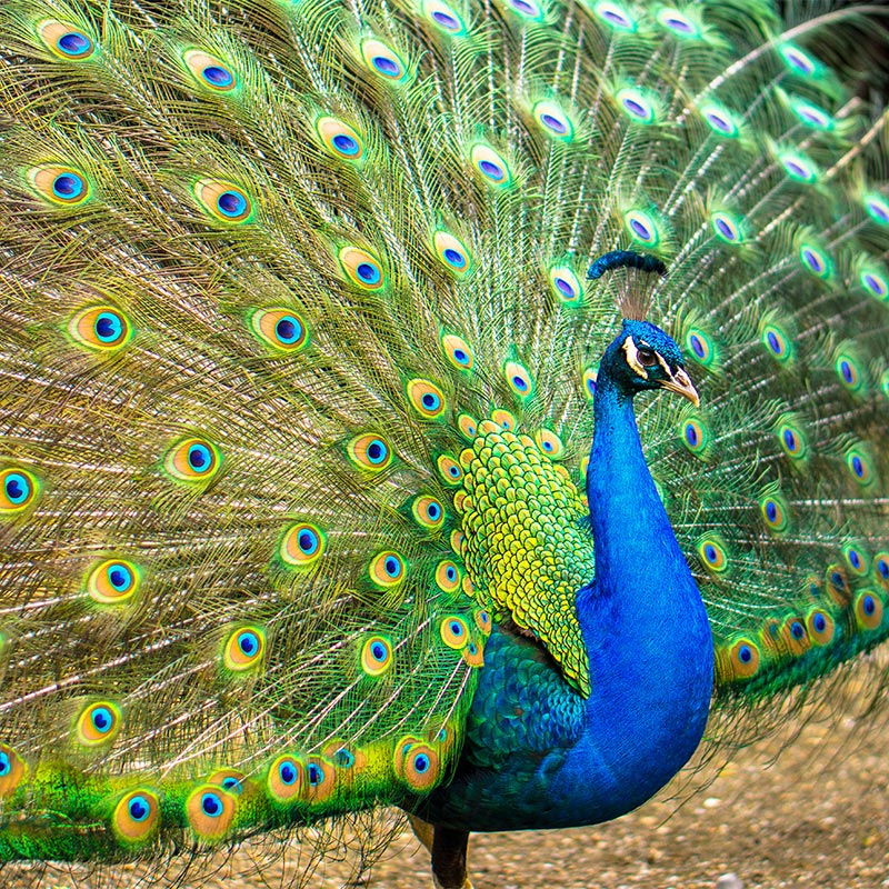
Structural color strikes again! It’s a pattern in nature to produce bright iridescent color by structural interactions with photons of light (called photonics) rather than pigment. Different wavelengths of light are absorbed or reflected producing particular colors and diffracting like a prism to produce iridescence. These colors are vibrant shimmering and rainbow-like, as seen in the peacock feather, butterfly scales, brightly colored fish, and gleaming insects.
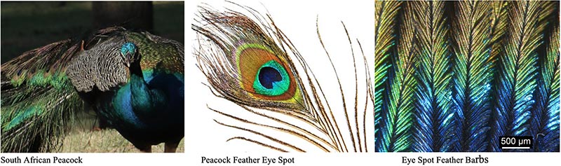
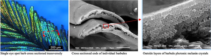
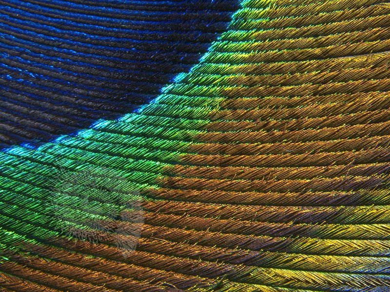
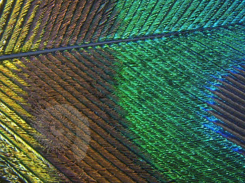
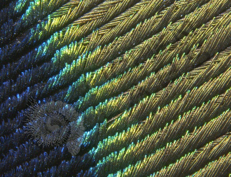
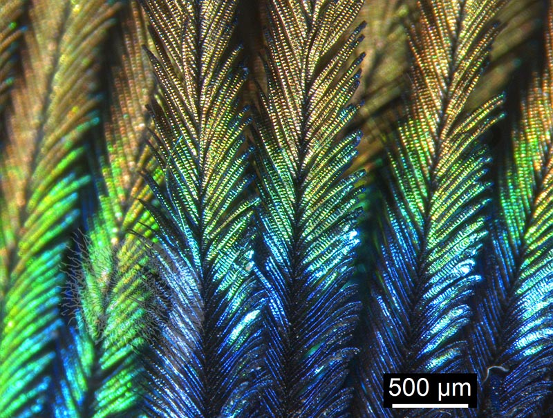
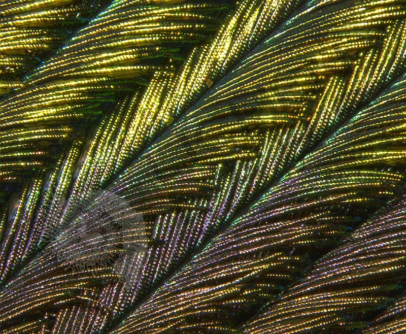
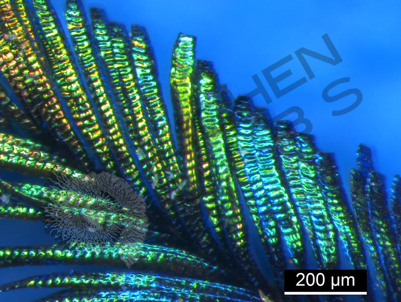
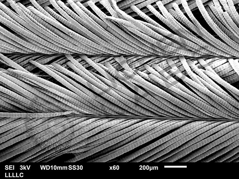
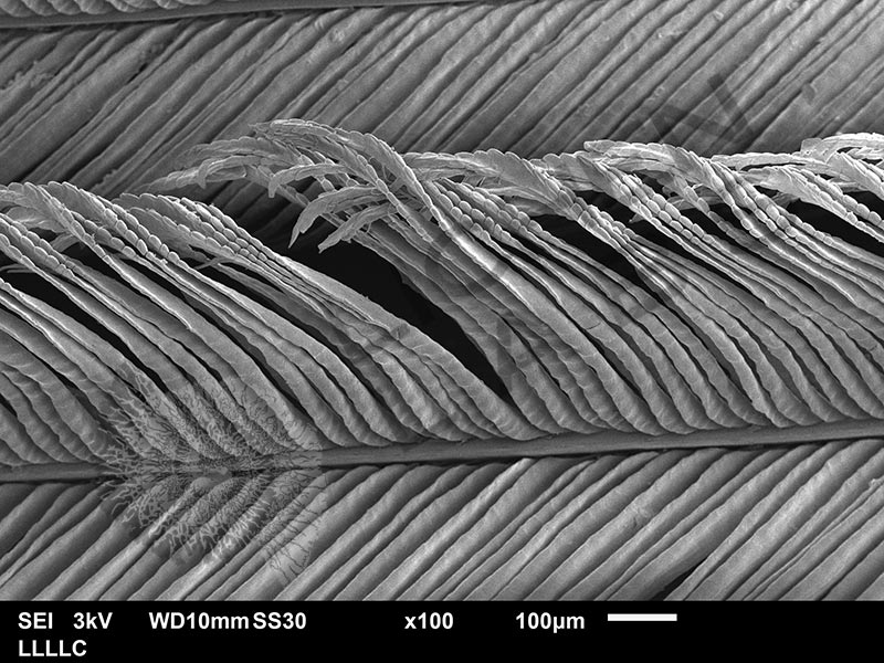
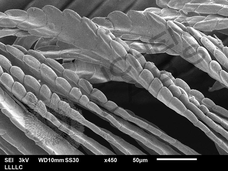
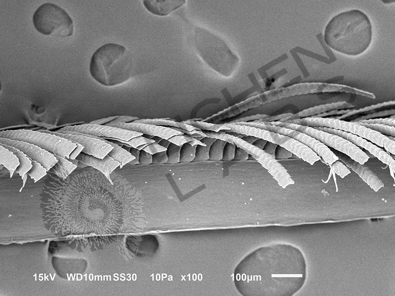
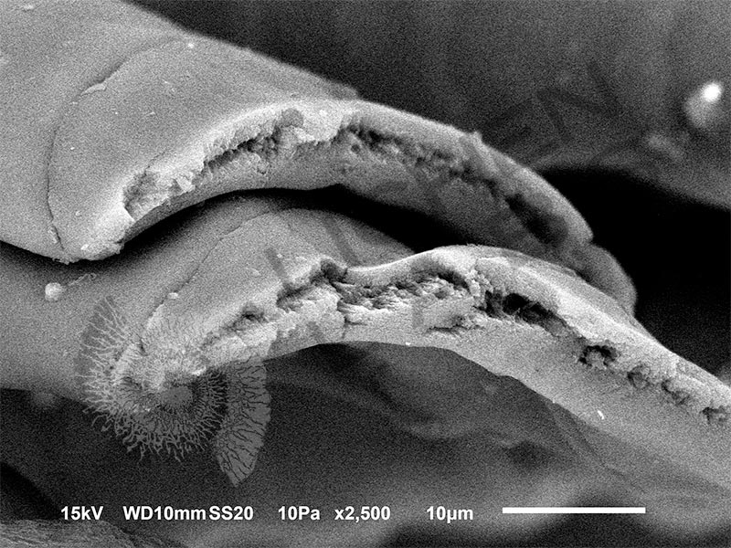
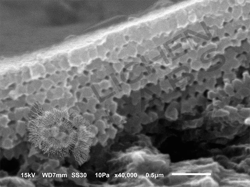
Above is a peacock seen in South Africa. The feather eye spot has many sub-feathers called barbs. Each barb has individual barbules extending from the fine shafts. To examine the internal structure, a razor blade was used to cut the barbules transversely. The end of the barbule was examined in the SEM. The outer structures contained rows of melanin photonic crystals. Melanin is the brown pigment found in our skin that protects us from the sun and also protects animals. In the case of the peacock, the melanin granules create optical interference that produces color. The curved structure of the barbules helps to diffuse the reflection, and the dark background the melanin pigment makes creates a more vivid interference color.
References:
https://en.wikipedia.org/wiki/Structural_coloration
https://pdfs.semanticscholar.org/8d3a/c5d1203292ccf71f736869d66aa94f6dd677.pdf
https://iopscience.iop.org/article/10.1088/1361-648X/aadc95
PURCHASE 5 OR MORE IMAGES AND GET 20% OFF YOUR ENTIRE ORDER!
Please contact us for custom images.
The image store is a collection of organisms that have been examined under a stereo light microscope (LM) and or scanning electron microscope (SEM). Each group of organisms has a short description and a longer more detailed description or story about the organism. Clicking on the product group shows the individual images. Each series takes the observer from macro to micro or nano on a particular organism, starting with a macro photographic image(s) for perspective, micro images taken by the light microscope, and most have micro to nano scanning electron microscope images. The SEM images will appear in black and white as a beam of electrons is used to illuminate the specimen rather than light. A few SEM images are colorized (lotus leaf). More information about the labeling and techniques used is below.
For the curious:
The light microscope images are labeled LM and a Z is included if it is a vertical composite of images effectively extending the depth of field or EDF of the microscope.
SEM images are labeled by the type of detector use:
SE (secondary electron)
LSE (Low vacuum secondary electron)
BES (backscattered electron shadow mode)
BEC (backscattered electron compositional mode)
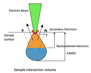
The SEM instrument works by producing a beam of electrons under a vacuum that interacts with the sample surface and subsurface producing different signals, as shown in the diagram at right. Secondary electrons, backscattered electrons and x-rays are detected using different instrument modes. In addition to morphological information to produce an image the SEM can determine elemental composition by energy dispersive x-ray spectroscopy (EDS).