Product Description
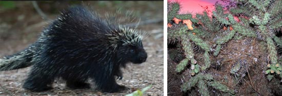
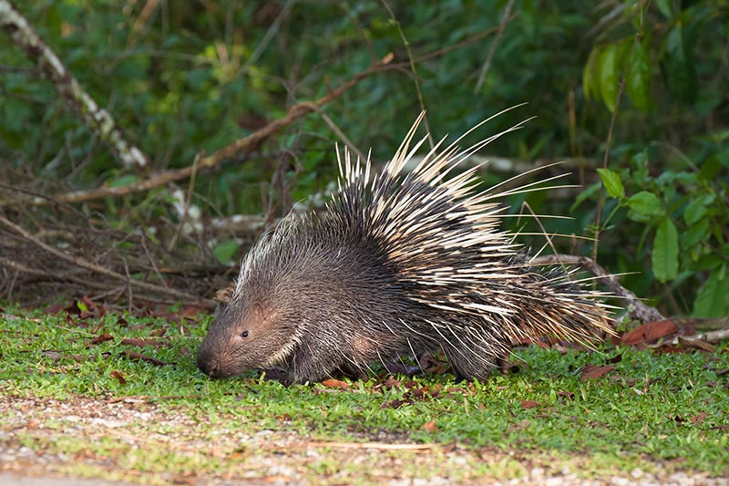
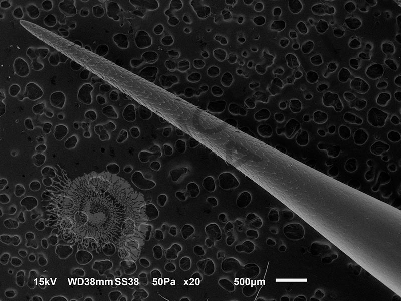
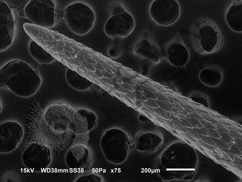
Porcupines have a spiny exterior to protect them from predators.

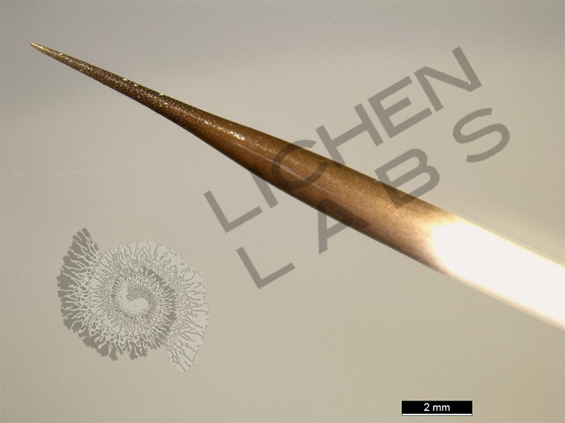
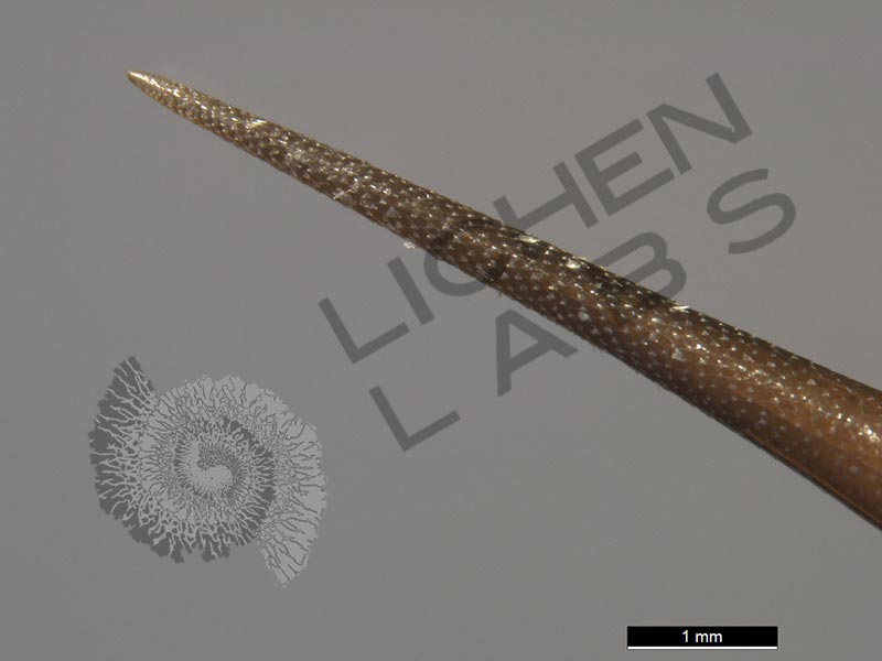
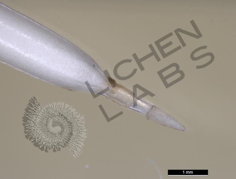
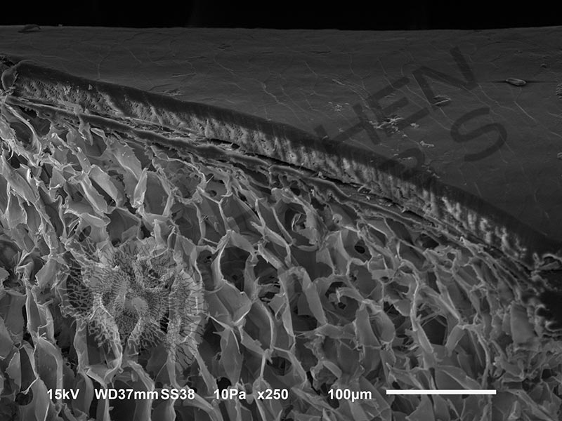
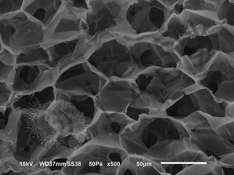
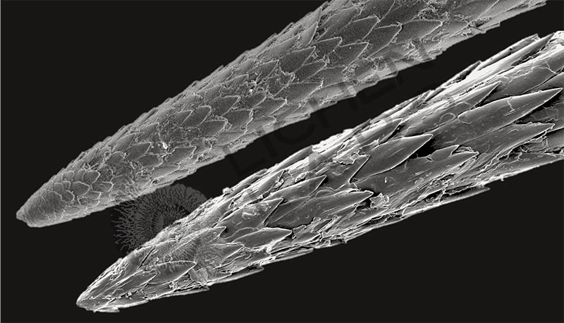
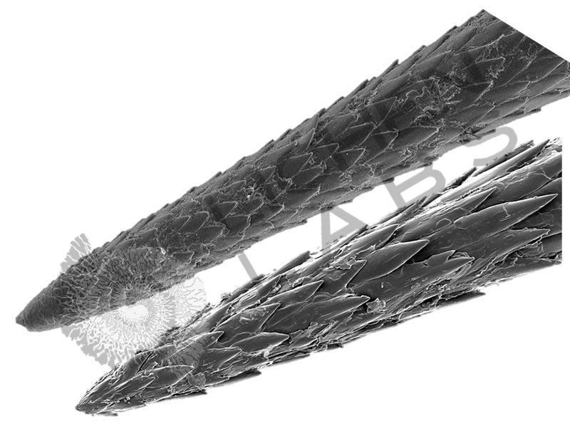
The porcupine quills slide in easily but have barbs in rows around their circumference. They penetrate deeply and are difficult to remove, leaving a lasting impression on would-be predators. The barbs also have a quick disconnect base that makes them appear to eject from the porcupine (see link below). The spines are light with a tough exterior coating and porous core.
http://www.naturalhistorymag.com/htmlsite/master.html

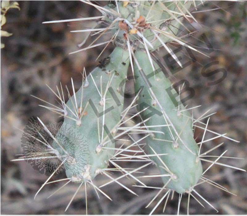
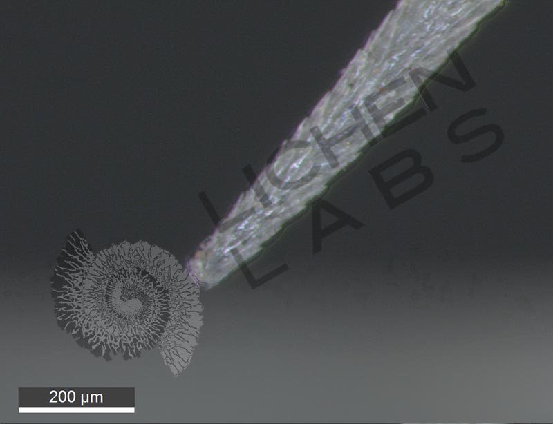
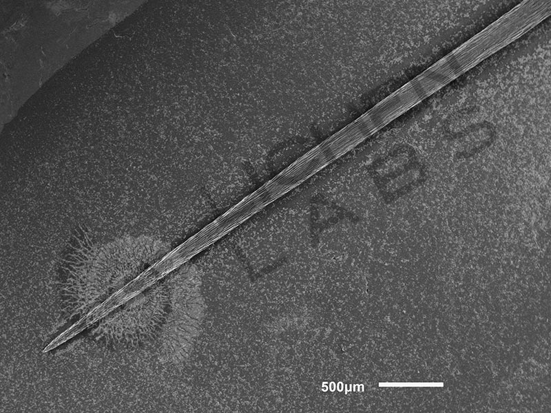
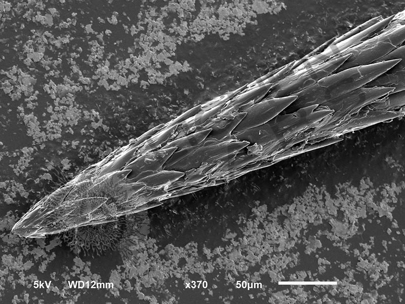
The very sharp spines of the Chain Fruit Cholla (Opuntia fulgida) have multitudes of fine barbs over the entire length. The barbs begin at 50 microns or millionths of a meter from the tip, which is approximately the width of a strand of hair. They are known as the “jumping cholla” because a slight brush by a passer-by results in extreme attachment. The joint on the cactus breaks off before the barbs pull out. These joint pieces can take root becoming vegetative propagules. It uses an attachment to desert animals as an additional dispersal tool and protection against attack.
https://eol.org/pages/478824
It is another pattern in nature with “jumping cholla” cactus spines and porcupine quills having similar structure and defense against predation.
Jeff Karp, a physician in Boston, designed a surgical staple emulating the barb structure of the porcupine quill.
https://www.cbsnews.com/news/biomimicry-turning-to-nature-for-technological-solutions/
https://www.pnas.org/content/109/52/21289
PURCHASE 5 OR MORE IMAGES AND GET 20% OFF YOUR ENTIRE ORDER!
Please contact us for custom images.
The image store is a collection of organisms that have been examined under a stereo light microscope (LM) and or scanning electron microscope (SEM). Each group of organisms has a short description and a longer more detailed description or story about the organism. Clicking on the product group shows the individual images. Each series takes the observer from macro to micro or nano on a particular organism, starting with a macro photographic image(s) for perspective, micro images taken by the light microscope, and most have micro to nano scanning electron microscope images. The SEM images will appear in black and white as a beam of electrons is used to illuminate the specimen rather than light. A few SEM images are colorized (lotus leaf). More information about the labeling and techniques used is below.
For the curious:
The light microscope images are labeled LM and a Z is included if it is a vertical composite of images effectively extending the depth of field or EDF of the microscope.
SEM images are labeled by the type of detector use:
SE (secondary electron)
LSE (Low vacuum secondary electron)
BES (backscattered electron shadow mode)
BEC (backscattered electron compositional mode)
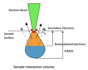
The SEM instrument works by producing a beam of electrons under a vacuum that interacts with the sample surface and subsurface producing different signals, as shown in the diagram at right. Secondary electrons, backscattered electrons and x-rays are detected using different instrument modes. In addition to morphological information to produce an image the SEM can determine elemental composition by energy dispersive x-ray spectroscopy (EDS).


