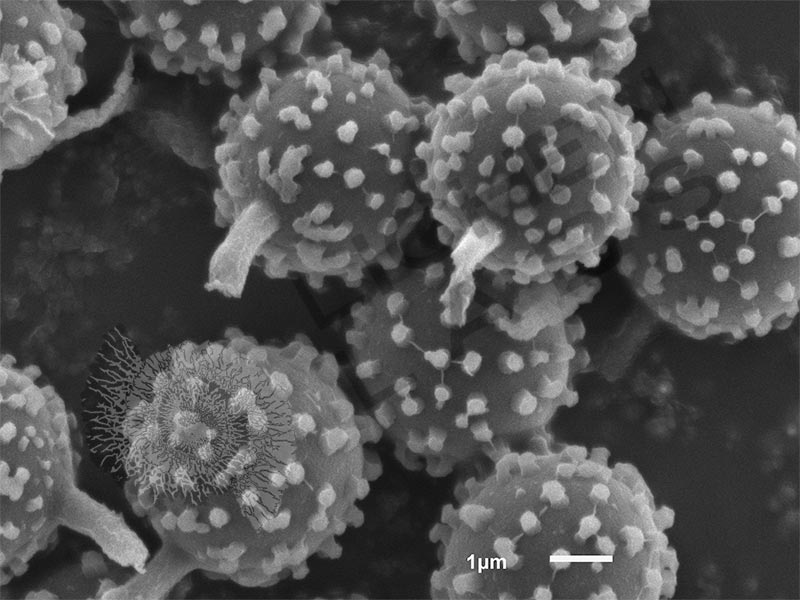
Puffball Spores
Price varies by image license.
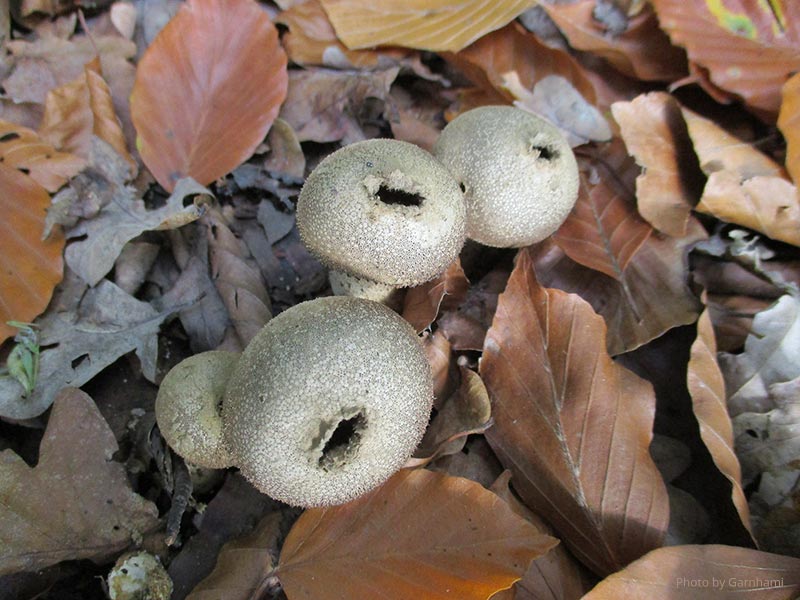
The common puffball mushroom (Lycoperdon perlatum) is cylindrical with a hole in the center where it releases its spores when it is bumped by animals or contacted by raindrops. The spores are so tiny and numerous they look like a puff of smoke as they exit.
Product Description
All multicellular fungi have two different morphological stages; vegetative and reproductive. Mycelium is the vegetative part of a fungus that consists of a tangle of branching slender threadlike structures called hyphae. The mycelium is typically underground or in rotting wood. Where it does the rotting. In the reproductive stage the mycelium sends up a mushroom technically called a “fruiting body” with the purpose of releasing reproductive cells called spores into the world.
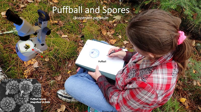
Girl near Ely, MN drawing a puffball in front of her left knee. One is also under a field microscope.
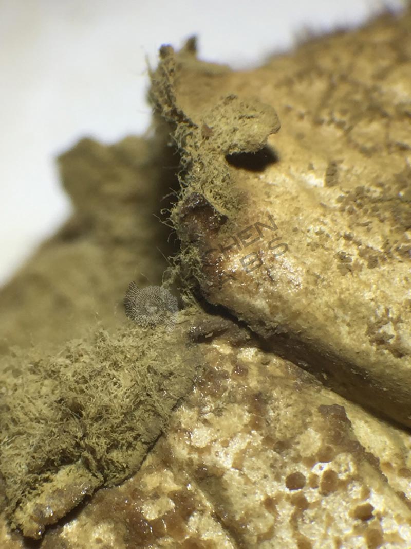
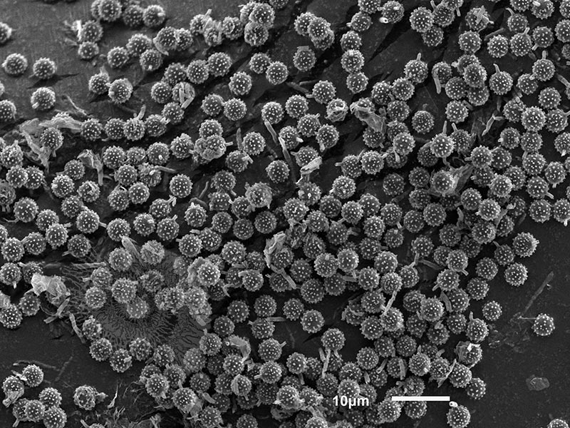
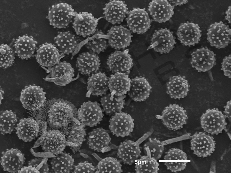
The common puffball is a type of fruiting body of a fungus that grows in rotting wood. The spores grow inside this type of mushroom within a spherical sac called the peridium. It has a single hole in the top. Once they mature, the mushroom becomes somewhat shrunken. With the slightest touch, it bursts open, releasing a cloud spores that look like a puff of smoke, which is why it gets its name. Each spore is connected to a turgid filament-like tube called a paraphyses that helps to provide the elastic prestress needed for spore ejection. Below are the puffball spores in 3 scanning electron microscope (SEM) images of increasing magnification. They are quite tiny ~ 4 mm (4 millionths of a meter) in diameter. A human hair ranges from 25 – 75 um in diameter. What’s left of the filament tube (paraphyses) assisting is clearly visible in the highest magnification taken at 10,000X.

References:
Field Guide: “Mushrooms of the Upper Midwest” by Teresa Marrone and Kathy Yerich, 2014
PURCHASE 5 OR MORE IMAGES AND GET 20% OFF YOUR ENTIRE ORDER!
Please contact us for custom images.
The image store is a collection of organisms that have been examined under a stereo light microscope (LM) and or scanning electron microscope (SEM). Each group of organisms has a short description and a longer more detailed description or story about the organism. Clicking on the product group shows the individual images. Each series takes the observer from macro to micro or nano on a particular organism, starting with a macro photographic image(s) for perspective, micro images taken by the light microscope, and most have micro to nano scanning electron microscope images. The SEM images will appear in black and white as a beam of electrons is used to illuminate the specimen rather than light. A few SEM images are colorized (lotus leaf). More information about the labeling and techniques used is below.
For the curious:
The light microscope images are labeled LM and a Z is included if it is a vertical composite of images effectively extending the depth of field or EDF of the microscope.
SEM images are labeled by the type of detector use:
SE (secondary electron)
LSE (Low vacuum secondary electron)
BES (backscattered electron shadow mode)
BEC (backscattered electron compositional mode)
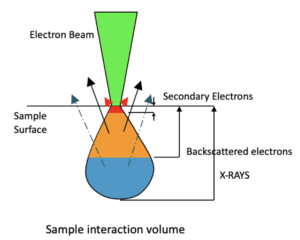
The SEM instrument works by producing a beam of electrons under a vacuum that interacts with the sample surface and subsurface producing different signals, as shown in the diagram at right. Secondary electrons, backscattered electrons and x-rays are detected using different instrument modes. In addition to morphological information to produce an image the SEM can determine elemental composition by energy dispersive x-ray spectroscopy (EDS).
Personal/Educational License:
For personal or educational use only. Images cannot be given or resold to others.
Commercial License:
For Profit or Non-profit business use within the organization, in presentations and publications with image credit. No resale of images.
An image license does not grant exclusive rights to an image. Lichen Labs retains the copywrite to all images.
