Product Description
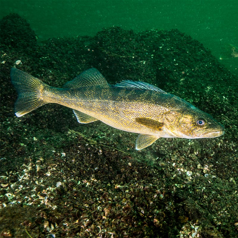
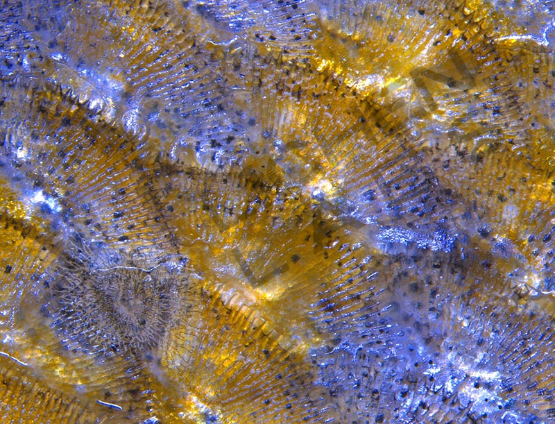
Walleye are the premier Minnesota game fish. I brought a Leica portable stereo microscope to our cabin on a northern Minnesota lake a few years ago. I wanted to show my daughter the lateral line and scales on a fish. After catching a walleye, I filleted and skinned it and immediately put the moist skin of the fish under the microscope.

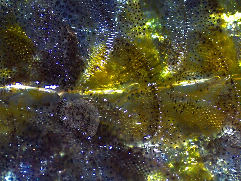
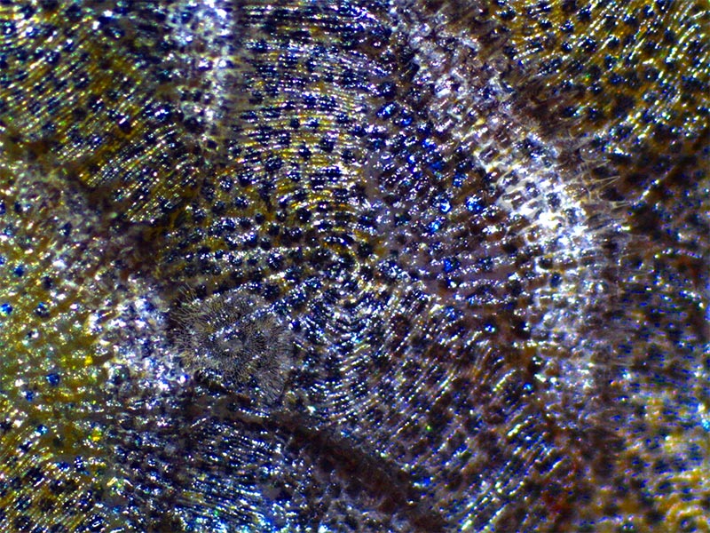
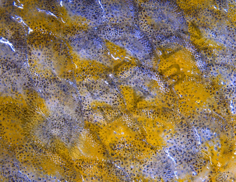
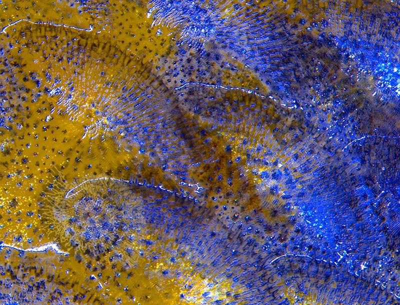
Blotches of vivid blue and gold patches of color reflected through the semi-clear scales were visible (above left), another example of structural color. Counting the rings (annuli) on a scale on a fish can be used to determine its age. Three pores of the lateral line system are visible on the side of the fish (above center). The two pores to the right have trapped air bubbles under the slime on the surface of the walleye. Those pores are openings to the lateral line canal that allow the fish to sense changes in pressure, movement, and vibration in the surrounding water (above right). Pressure changes deflect the neuromast cells on the tip of the hair cilium that is connected in bundles to its nervous system.
References
https://www.wikiwand.com/en/Walleye
Lateral Line System of Fish
https://onlinelibrary.wiley.com/doi/abs/10.1111/j.1749-4877.2008.00131.x
https://en.wikipedia.org/wiki/Lateral_line
PURCHASE 5 OR MORE IMAGES AND GET 20% OFF YOUR ENTIRE ORDER!
Please contact us for custom images.
The image store is a collection of organisms that have been examined under a stereo light microscope (LM) and or scanning electron microscope (SEM). Each group of organisms has a short description and a longer more detailed description or story about the organism. Clicking on the product group shows the individual images. Each series takes the observer from macro to micro or nano on a particular organism, starting with a macro photographic image(s) for perspective, micro images taken by the light microscope, and most have micro to nano scanning electron microscope images. The SEM images will appear in black and white as a beam of electrons is used to illuminate the specimen rather than light. A few SEM images are colorized (lotus leaf). More information about the labeling and techniques used is below.
For the curious:
The light microscope images are labeled LM and a Z is included if it is a vertical composite of images effectively extending the depth of field or EDF of the microscope.
SEM images are labeled by the type of detector use:
SE (secondary electron)
LSE (Low vacuum secondary electron)
BES (backscattered electron shadow mode)
BEC (backscattered electron compositional mode)
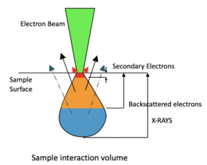
The SEM instrument works by producing a beam of electrons under a vacuum that interacts with the sample surface and subsurface producing different signals, as shown in the diagram at right. Secondary electrons, backscattered electrons and x-rays are detected using different instrument modes. In addition to morphological information to produce an image the SEM can determine elemental composition by energy dispersive x-ray spectroscopy (EDS).